Orthopedics
Acute comminuted intertrochanteric hip fracture
DOI: https://doi.org/10.21980/J8QK9CHistory of present illness: A 69-year-old male presented to the emergency department (ED) with left hip pain after he was rear-ended on his bicycle by a vehicle traveling 10-15 miles per hour. He had normal vital signs. On exam, his left lower extremity was externally rotated and shortened with trochanteric point tenderness. His pelvis was stable. His lower extremity compartments
Rare Rapidly Growing Thumb Lesion in a 12-Year-Old Male
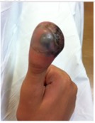
DOI: https://doi.org/10.21980/J8B92JHistory of present illness: A 12-year-old male presented to the emergency department with right thumb pain and a mass for four months (see images). He denied fevers, chills, change in appetite, or fatigue. He noted that the lesion was growing and “bleeds easily if bumped.” He denied any trauma to the thumb, except “hitting it” months ago while in football
Biceps Tendon Rupture
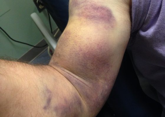
DOI: https://doi.org/10.21980/J8RP8BPhysical exam was significant for ecchymosis and mild swelling of the right bicep. When the right arm was flexed at the elbow, a prominent mass was visible and palpable over the right bicep. Right upper extremity strength was 4/5 with flexion at the elbow.
Bilateral Hip Dislocation in Unrestrained Driver
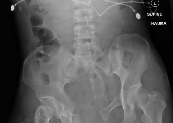
DOI: https://doi.org/10.21980/J8HD0CThe initial radiograph of the pelvis revealed bilateral hip dislocations. Small bony fragments were noted in the right hip joint, suggestive of an underlying fracture. The sacroiliac joints and the pelvic ring were intact. In the emergency department, bilateral hip reductions were performed using the Captain Morgan technique.1 The post-reduction film showed reduction of the bilateral hip dislocations with extensive comminuted and displaced fractures of the right and left acetabula.
Open Book Pelvic Fracture
DOI: https://doi.org/10.21980/J8CK7HThe initial radiograph of the pelvis shows an open-book pelvic fracture deformity with pubic symphyseal dislocation, left greater than right sacroiliac diastases, and fractures of the left superior and inferior pubic rami, right inferior pubic ramus, and left acetabular anterior column. The additional inlet and outlet radiographs of the pelvis after application of a pelvic binder also show an open book fracture with significant improvement of the widened pubic symphysis.
Subcutaneous Emphysema in Non-Necrotizing Soft Tissue Injury
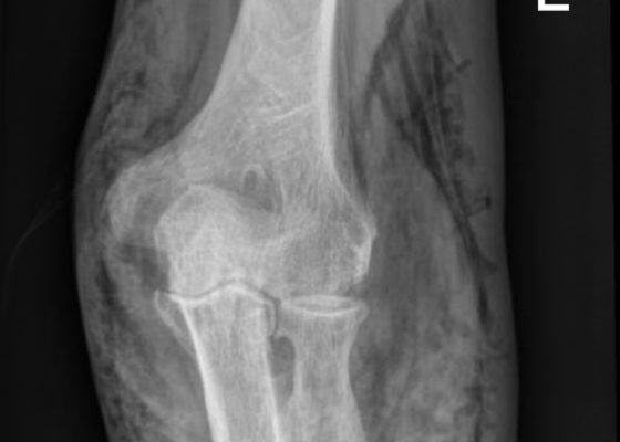
DOI: https://doi.org/10.21980/J8432MX-Rays of the elbow revealed diffuse striated lucencies throughout the soft tissue, consistent with extensive subcutaneous air throughout the superficial and deep tissues. There was no evidence of a fracture.
Acetabular Fracture
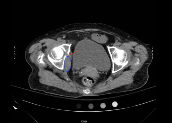
DOI: https://doi.org/10.21980/J8BK8KThe non-contrast CT images show a minimally displaced comminuted fracture of the right acetabulum involving the acetabular roof, medial and anterior walls (red arrows), with associated obturator muscle hematoma (blue oval).
A Simulation Model for Extensor Tendon Repair
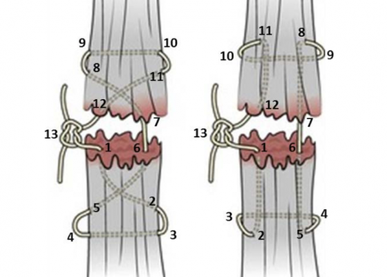
DOI: https://doi.org/10.21980/J8VS7XBy the end of this educational session, the learner will be able to: 1) List the indications for extensor tendon repair in the emergency department, 2) recognize the indications for referral to orthopedic or hand surgery, 3) list the risks and benefits of emergency department extensor tendon repair, 4) perform an appropriate physical examination for a patient with a potential extensor tendon laceration, 5) list the maximum time limit of tourniquet application for this procedure, 6) list the materials needed for extensor tendon repair in the emergency department, 7) successfully repair a completely severed extensor tendon using four different techniques: horizontal mattress, figure of eight, modified Kessler and modified Bunnell, and 8) describe the appropriate splinting of a repaired extensor tendon.


