• Registration
  • Login
JETem
  • Home
  • About
    • Aim and Scope
    • Our Team
    • Editorial Board
    • FAQ
  • Issues
    • Current Issue
    • Ahead of Print
    • Past Issues
  • Visual EM
    • Latest Visual EM
    • Search Visual EM
    • Thumbnail Library
  • For Authors
    • Instructions for Authors
    • Submit to JETem
    • Photo Consent
    • Policies
      • Peer Review Policy
      • Copyright Policy
      • Editorial Policy, Ethics and Responsibilities
      • Conflicts of Interest & Informed Consent
      • Open Access Policy
  • For Reviewers
    • Instructions for JETem Reviewers
    • Interested in Being a JETem Reviewer?
  • Topic
    • Abdominal / Gastroenterology
    • Administration
    • Board Review
    • Cardiology / Vascular
    • Clinical Informatics, Telehealth and Technology
    • Dermatology
    • EMS
    • Endocrine
    • ENT
    • Faculty Development
    • Genitourinary
    • Geriatrics
    • Hematology / Oncology
    • Infectious Disease
    • Miscellaneous
    • Neurology
    • Ob / Gyn
    • Ophthalmology
    • Orthopedics
    • Pediatrics
    • Pharmacology
    • Procedures
    • Psychiatry
    • Renal / Electrolytes
    • Respiratory
    • Social Determinants of Health
    • Toxicology
    • Trauma
    • Ultrasound
    • Urology
    • Wellness
    • Wilderness
  • Modality
    • Curricula
    • Innovations
    • Lectures
    • Oral Boards
      • Structured Interview
      • Communication Case
    • Podcasts
    • Simulation
    • Small Group Learning
    • Team Based Learning
    • Visual EM
  • Contact Us

Orthopedics

Creative Commons images

Biceps Tendon Rupture

Daniel M Polvino, MD* and Grant Wei, MD*

DOI: https://doi.org/10.21980/J8RP8B Issue 3:1 No ratings yet.
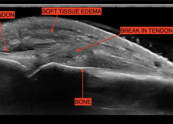
Physical exam was significant for ecchymosis and mild swelling of the right bicep. When the right arm was flexed at the elbow, a prominent mass was visible and palpable over the right bicep. Right upper extremity strength was 4/5 with flexion at the elbow.
OrthopedicsVisual EM
Creative Commons images

Bilateral Hip Dislocation in Unrestrained Driver

Samer Assaf, MD* and Ghadi Ghanem*

DOI: https://doi.org/10.21980/J8HD0C Issue 2:4 No ratings yet.
The initial radiograph of the pelvis revealed bilateral hip dislocations. Small bony fragments were noted in the right hip joint, suggestive of an underlying fracture. The sacroiliac joints and the pelvic ring were intact. In the emergency department, bilateral hip reductions were performed using the Captain Morgan technique.1 The post-reduction film showed reduction of the bilateral hip dislocations with extensive comminuted and displaced fractures of the right and left acetabula.
OrthopedicsVisual EM
Creative Commons images

Open Book Pelvic Fracture

Samer Assaf, MD* and Ghadi Ghanem*

DOI: https://doi.org/10.21980/J8CK7H Issue 2:3 No ratings yet.
The initial radiograph of the pelvis shows an open-book pelvic fracture deformity with pubic symphyseal dislocation, left greater than right sacroiliac diastases, and fractures of the left superior and inferior pubic rami, right inferior pubic ramus, and left acetabular anterior column. The additional inlet and outlet radiographs of the pelvis after application of a pelvic binder also show an open book fracture with significant improvement of the widened pubic symphysis.
OrthopedicsTraumaVisual EM
Creative Commons images

Subcutaneous Emphysema in Non-Necrotizing Soft Tissue Injury

Hamid Ehsani-Nia, MS, DO* and Lauren Sylwanowicz MD ^

DOI: https://doi.org/10.21980/J8432M Issue 2:4 No ratings yet.
X-Rays of the elbow revealed diffuse striated lucencies throughout the soft tissue, consistent with extensive subcutaneous air throughout the superficial and deep tissues. There was no evidence of a fracture.
Infectious DiseaseOrthopedicsVisual EM
Creative Commons images

Acetabular Fracture

Chad Correa* and Sari Lahham, MD^

DOI: https://doi.org/10.21980/J8BK8K Issue 2:4 No ratings yet.
The non-contrast CT images show a minimally displaced comminuted fracture of the right acetabulum involving the acetabular roof, medial and anterior walls (red arrows), with associated obturator muscle hematoma (blue oval).
OrthopedicsVisual EM
Creative Commons images

A Simulation Model for Extensor Tendon Repair

Elizabeth Aronstam, DO* and David T Overton, MD*

DOI: https://doi.org/10.21980/J8VS7X Issue 2:3 No ratings yet.
By the end of this educational session, the learner will be able to: 1) List the indications for extensor tendon repair in the emergency department, 2) recognize the indications for referral to orthopedic or hand surgery, 3) list the risks and benefits of emergency department extensor tendon repair, 4) perform an appropriate physical examination for a patient with a potential extensor tendon laceration, 5) list the maximum time limit of tourniquet application for this procedure, 6) list the materials needed for extensor tendon repair in the emergency department, 7) successfully repair a completely severed extensor tendon using four different techniques: horizontal mattress, figure of eight, modified Kessler and modified Bunnell, and 8) describe the appropriate splinting of a repaired extensor tendon.
ProceduresInnovationsOrthopedics
Creative Commons images

Introducing point-of-care ultrasound through competency-based simulation education using a fractured chicken bone model

Nathan L Haas, MD,* Elise Hart, MD,† Mary RC Haas, MD,* and Trent Reed, DO^

DOI: https://doi.org/10.21980/J8GG95 Issue 2:3 No ratings yet.
To introduce medical students to PoCUS with an inexpensive, reproducible, and educationally effective model using fractured chicken bones set in gelatin, and to assess medical students’ abilities to identify simulated long-bone fractures using PoCUS.
UltrasoundInnovationsOrthopedics
Creative Commons images

Supracondylar Fracture

Jessica Andrusaitis, BS, MS* and Ben Feldman, MD*

DOI: https://doi.org/10.21980/J8492P Issue 2:3 No ratings yet.
History of present illness: A 15-year-old male presented to the emergency department with right elbow pain after falling off a skateboard.  The patient denied a decrease in strength or sensation but did endorse paresthesias to his hand.  On exam, the patient had an obvious deformity of his right elbow with tenderness to palpation and decreased range of motion at the
OrthopedicsVisual EM
Creative Commons images

Galeazzi Fracture

Reid Honda, MD*

DOI: https://doi.org/10.21980/J8HS39 Issue 2:2 No ratings yet.
The X-ray showed an acute comminuted fracture of the distal diaphysis of the radius with disruption of the distal radioulnar joint, consistent with a Galeazzi fracture. The patient was then splinted and taken for operative reduction and internal fixation the following day.
OrthopedicsVisual EM

A Formalized Three-Year Emergency Medicine Residency Musculoskeletal Emergencies Curriculum

Andrew King, MD, FACEP*, Jeffrey Yu^, Mark J Conroy, MD*, Robert Cooper, MD*, Jennifer Mitzman, MD*, Colin Kaide, MD, FACEP* Sarah Greenberger, MD*, Sorabh Khandelwal, MD* and Michael Barrie, MD*

DOI: https://doi.org/10.21980/J8RG6HIssue 2:1 No ratings yet.
Resident learners will master the diagnosis and management of emergent musculoskeletal conditions including fractures/dislocations, soft tissue injuries, compartment syndrome, joint complaints, infections, and complex injuries.
OrthopedicsCurricula
«‹5›»
Page 5 of 7

JETem is an online, open access, peer-reviewed, journal-repository for EM educators. We are PMC Indexed.

Most Viewed

  • The Silent Saboteur: Teaching the Clinical Implications of Occult Hypoxemia & Social Determinants of Health via a Pulmonary Embolism Case
  • Diabetic Ketoacidosis and Necrotizing Soft Tissue Infection
  • My Broken Heart
  • Stabilization of Cardiogenic Shock for Critical Care Transport, a Simulation
  • Innovative Ultrasound-Guided Erector Spinae Plane Nerve Block Model for Training Emergency Medicine Physicians

Visit Our Collaborators

Creative Commons Licence
This work is licensed under a Creative Commons Attribution 4.0 International License.

About

Education

Learners should benefit from active learning. JETem accepts submissions of team-based learning, small group learning, simulation, podcasts, lectures, innovations, curricula, question sets, and visualEM.

Scholarship

We believe educators should advance through the scholarship of their educational work. JETem gives educators the opportunity to publish scholarly academic work so that it may be widely distributed, thereby increasing the significance of their results.

Links

  • Home
  • Aim and Scope
  • Current Issue
  • For Reviewers
  • Instructions for Authors
  • Contact Us

Newsletter

Sign up to receive updates from JETem regarding newly published issues and findings.

Copyright Creative Commons Attribution 4.0 International