
Orthopedics


Case Report of the Unusual Presentation of Stridor in an Elderly Patient Following a Cervical Fracture
DOI: https://doi.org/10.21980/J8V926The cervical CT was significant for a transverse fracture through the C4 vertebral body (see red arrow), lateral facet (green arrow), spinous process (blue arrow), and right lamina (purple arrow) as well as surrounding edema and retropharyngeal thickening (yellow line), best appreciated on sagittal view.

Digital Nerve Block for the Reduction of a Proximal Phalanx Fracture of the Foot – a Case Report
DOI: https://doi.org/10.21980/J8KS8TPlain film of the right foot showed evidence of an oblique fracture of the body of the proximal 4th phalanx (image 2). No other acute traumatic injuries noted in the rest of the bones and joints of the right foot. After performing a digital block of the toe and reduction, repeat imaging showed evidence of successful reduction with anatomic alignment and redemonstration of the fracture line (image 3).
Open Subtalar Dislocation
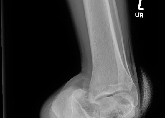
DOI: https://doi.org/10.21980/J87P8PX-ray of the left ankle revealed a complete dislocation of the subtalar joint with medial dislocation of the calcaneus (outlined in orange) relative to the talus (outlined in red) with subcutaneous air noted in the lateral soft tissues (blue arrows in Figure 1). The talonavicular joint has also been disrupted (navicular outlined in blue). There was no evidence of fracture. Post-reduction computed tomography of the left lower extremity confirmed no evidence of associated fracture.

Ultrasonographic Findings of Acute Achilles Tendon Rupture
DOI: https://doi.org/10.21980/J8063SThe ultrasound video clip shows a longitudinal view of the AT during a dynamic exam. While the patient’s foot is gently passively dorsi/plantar flexed, the video demonstrates first a normal Achilles tendon (from the unaffected extremity) without disruption (highlighted by a yellow bracket on screen left). Then it shows an abnormal tendon (the patient’s affected side) with disruption of the typical linear tendon fibers (red arrow identifies area of rupture). Dynamic testing shows the movement of the distal stump of the AT is evident adjacent to hypoechoic fluid that is reactive edema or blood from the acute rupture.

Medial Subtalar Dislocation: A Case Report
DOI: https://doi.org/10.21980/J8QP9DOn examination, the patient had a significant deformity to his left foot and ankle. His left foot was pointed medially. His distal left tibia and fibula were visible and palpable upon inspection, with the overlying skin completely intact. There was no neurovascular compromise of the foot. X-ray demonstrated a subtalar dislocation of the left ankle (green arrow) and significant widening of the tibiotalar joint space (yellow arrow). There was associated soft tissue swelling but no acute displaced fractures were identified.

Classic Slipped Capital Femoral Epiphysis: A Case Report
DOI: https://doi.org/10.21980/J8BD16The pelvis X-ray demonstrates a widened right capital femoral epiphysis (more than 2 mm) that is typical of a slipped capital femoral epiphysis (SCFE).1 The yellow highlight outlines this area of widening.
The classic Klein’s line (orange lines) is often inaccurate and even difficult to draw with certainty.1 Nevertheless, in this X-ray, one has a sense that the right capital femoral epiphysis does not align with the femoral neck in the same way as it does on the left side, suggesting slippage.
Point-Of-Care Ultrasound for the Diagnosis of Extensor Tenosynovitis
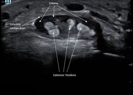
DOI: https://doi.org/10.21980/J8Q050Point-of-care ultrasound of the dorsal aspect of the left hand reveals a heterogenous hypoechoic fluid collection surrounding the extensor tendons (axial view) within the retinaculum consistent with edema. Longitudinal view shows anechoic fluid within the tenosynovium which is located between the anisotropic extensor tendon and linear hyperechoic synovial sheath. Longitudinal view also shows some cobblestoning, or tissue edema, superficial to the anisotropic extensor tendon. The patient’s contralateral right dorsal hand was scanned in a longitudinal view and shows no cobblestoning or hypoechoic fluid under the synovial sheath. The patient was diagnosed with tenosynovitis, and started on intravenous antibiotics.

Asymptomatic CT Iodinated Contrast Extravasation of the Upper Extremity
DOI: https://doi.org/10.21980/J8VK87The two radiographs demonstrate extravasation of radiopaque iodinated contrast in the lower left upper extremity with most seen in the left antecubital fossa and left proximal forearm. Extravasation is seen in the subcutaneous and subfascial tissue.