• Registration
  • Login
JETem
  • Home
  • About
    • Aim and Scope
    • Our Team
    • Editorial Board
    • FAQ
  • Issues
    • Current Issue
    • Ahead of Print
    • Past Issues
  • Visual EM
    • Latest Visual EM
    • Search Visual EM
    • Thumbnail Library
  • For Authors
    • Instructions for Authors
    • Submit to JETem
    • Photo Consent
    • Policies
      • Peer Review Policy
      • Copyright Policy
      • Editorial Policy, Ethics and Responsibilities
      • Conflicts of Interest & Informed Consent
      • Open Access Policy
  • For Reviewers
    • Instructions for JETem Reviewers
    • Interested in Being a JETem Reviewer?
  • Topic
    • Abdominal / Gastroenterology
    • Administration
    • Board Review
    • Cardiology / Vascular
    • Clinical Informatics, Telehealth and Technology
    • Dermatology
    • EMS
    • Endocrine
    • ENT
    • Faculty Development
    • Genitourinary
    • Geriatrics
    • Hematology / Oncology
    • Infectious Disease
    • Miscellaneous
    • Neurology
    • Ob / Gyn
    • Ophthalmology
    • Orthopedics
    • Pediatrics
    • Pharmacology
    • Procedures
    • Psychiatry
    • Renal / Electrolytes
    • Respiratory
    • Social Determinants of Health
    • Toxicology
    • Trauma
    • Ultrasound
    • Urology
    • Wellness
    • Wilderness
  • Modality
    • Curricula
    • Innovations
    • Lectures
    • Oral Boards
      • Structured Interview
      • Communication Case
    • Podcasts
    • Simulation
    • Small Group Learning
    • Team Based Learning
    • Visual EM
  • Contact Us

Orthopedics

Creative Commons images

Ultrasonographic Findings of Acute Achilles Tendon Rupture

Charles Craig Rudy, MD*^, John A Thompson, MD* and Rachel R Bengtzen, MD*^

DOI: https://doi.org/10.21980/J8063S Issue 4:4 No ratings yet.
The ultrasound video clip shows a longitudinal view of the AT during a dynamic exam. While the patient’s foot is gently passively dorsi/plantar flexed, the video demonstrates first a normal Achilles tendon (from the unaffected extremity) without disruption (highlighted by a yellow bracket on screen left).  Then it shows an abnormal tendon (the patient’s affected side) with disruption of the typical linear tendon fibers (red arrow identifies area of rupture). Dynamic testing shows the movement of the distal stump of the AT is evident adjacent to hypoechoic fluid that is reactive edema or blood from the acute rupture. 
OrthopedicsVisual EM
Creative Commons images

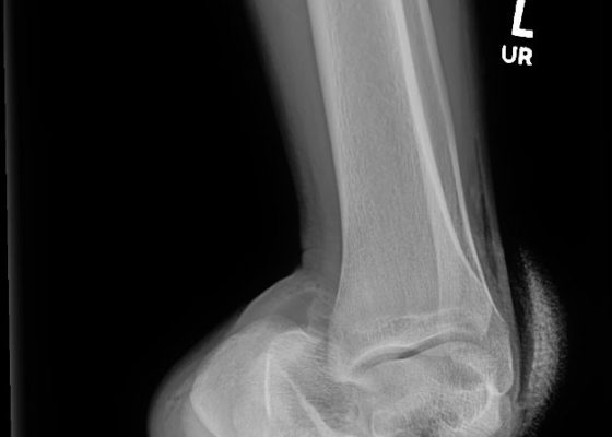
Medial Subtalar Dislocation: A Case Report

Claire Thomas, MD*  and Annasha Vyas, BS*

DOI: https://doi.org/10.21980/J8QP9D Issue 4:4 No ratings yet.
On examination, the patient had a significant deformity to his left foot and ankle. His left foot was pointed medially. His distal left tibia and fibula were visible and palpable upon inspection, with the overlying skin completely intact. There was no neurovascular compromise of the foot. X-ray demonstrated a subtalar dislocation of the left ankle (green arrow) and significant widening of the tibiotalar joint space (yellow arrow). There was associated soft tissue swelling but no acute displaced fractures were identified.
OrthopedicsVisual EM
Creative Commons images

Classic Slipped Capital Femoral Epiphysis: A Case Report

James Webley, MD*

DOI: https://doi.org/10.21980/J8BD16 Issue 4:4 No ratings yet.
The pelvis X-ray demonstrates a widened right capital femoral epiphysis (more than 2 mm) that is typical of a slipped capital femoral epiphysis (SCFE).1 The yellow highlight outlines this area of widening. The classic Klein’s line (orange lines) is often inaccurate and even difficult to draw with certainty.1 Nevertheless, in this X-ray, one has a sense that the right capital femoral epiphysis does not align with the femoral neck in the same way as it does on the left side, suggesting slippage.
OrthopedicsVisual EM
Creative Commons images

Point-Of-Care Ultrasound for the Diagnosis of Extensor Tenosynovitis

James A Frank, MD*, Joshua Lupton, MD* and Bryson Hicks, MD*

DOI: https://doi.org/10.21980/J8Q050Issue 4:3 No ratings yet.
Point-of-care ultrasound of the dorsal aspect of the left hand reveals a heterogenous hypoechoic fluid collection surrounding the extensor tendons (axial view) within the retinaculum consistent with edema. Longitudinal view shows anechoic fluid within the tenosynovium which is located between the anisotropic extensor tendon and linear hyperechoic synovial sheath. Longitudinal view also shows some cobblestoning, or tissue edema, superficial to the anisotropic extensor tendon. The patient’s contralateral right dorsal hand was scanned in a longitudinal view and shows no cobblestoning or hypoechoic fluid under the synovial sheath. The patient was diagnosed with tenosynovitis, and started on intravenous antibiotics.
OrthopedicsInfectious DiseaseUltrasoundVisual EM
Creative Commons images

Asymptomatic CT Iodinated Contrast Extravasation of the Upper Extremity

Eric Liao, MD* and John Costumbrado, MD, MPH*

DOI: https://doi.org/10.21980/J8VK87 Issue 4:3 No ratings yet.
The two radiographs demonstrate extravasation of radiopaque iodinated contrast in the lower left upper extremity with most seen in the left antecubital fossa and left proximal forearm. Extravasation is seen in the subcutaneous and subfascial tissue.
Visual EMOrthopedics
Creative Commons images

Open Fracture of the Patella

Zara Khan, BS*, Devan Pandya, MD^ and John Costumbrado, MD, MPH^

DOI: https://doi.org/10.21980/J8BK9Z Issue 4:3 No ratings yet.
X-ray of the right knee showed evidence of an acute comminuted fracture of the patella (red arrows) with a suprapatellar joint effusion with gas (blue arrow). There was no evidence of joint dislocation or other osseous lesions.
OrthopedicsVisual EM
Creative Commons images

Diagnosis and Treatment of an Anterior Shoulder Dislocation with Bedside Ultrasound

Mary Rometti, MD*, Michael Rohinton Mirza, MD* and Christopher Bryczkowski, MD*

DOI: https://doi.org/10.21980/J8Z924Issue 4:3 No ratings yet.
Bedside ultrasound with the transducer placed on the posterior right shoulder revealed an anterior dislocation of the right humerus. This is evident by displacement of the humeral head further away from the posteriorly placed ultrasound transducer, and appears deep to the glenoid cavity.  In a posterior shoulder dislocation, the humeral head would appear closer to the transducer (and the near field of the ultrasound image) than the glenoid. Note that a hypoechoic, heterogeneous fluid collection is within the joint space, compatible with a hematoma. A right shoulder X-ray confirmed the anterior dislocation with no evidence of fracture.  Under direct ultrasound guidance the glenohumeral joint space was injected with 10 mL of 2% lidocaine as an intraarticular anesthetic block. The right shoulder was reduced using continual traction.  Post-reduction ultrasound demonstrated a successful shoulder reduction, depicted by the humeral head being relocated to its anatomical location, adjacent to the glenoid cavity, as noted on the ultrasound image.  A hematoma remains present within the joint space. Successful shoulder reduction was further confirmed by X-ray. The patient’s arm was placed in a sling and she was discharged home with orthopedics follow-up.
OrthopedicsVisual EM
Creative Commons images

Pectoralis Muscle Tendon Rupture

Wyatt Verplaetse, MD* and Gregory Podolej, MD*

DOI: https://doi.org/10.21980/J81D01Issue 4:2 No ratings yet.
There is a noticeable difference in appearance and location of maximal prominence of the right pectoral muscle with arms outstretched (image 1). This is accentuated by having the patient perform an isometric arm press. (image 2).There is absence of the anterior axillary fold with adduction against resistance. The stump of the pectoralis muscle was palpated along his armpit.  He otherwise has full range of motion in the shoulder with minimal pain.
Visual EMOrthopedics
Creative Commons images

Pediatric Sedation for Forearm Fracture

Nichole Niknafs, DO* and Alisa Wray, MD, MAEd^

DOI: https://doi.org/10.21980/J8CS7K Issue 4:1 No ratings yet.
At the end of this simulation, participants will: 1) review options for pain control in pediatric patients, 2) perform a pre-sedation history and physical exam, 3) review the indications and contraindications for pediatric moderate sedation, 4) understand components of consent, and get consent from the patient’s parent, 5) list medication options for moderate sedation in a pediatric patient and review their appropriate doses, indications, contraindications, and side effects, 6) discuss management of moderate sedation complications, and 7) review criteria for discharging a patient after sedation. 
PediatricsOrthopedicsProcedures
Creative Commons images

Bilateral Shoulder Dislocation after Ski Injury

Alaina Rajagopal, PhD, MD*, Brian Knight, BS* and Lance Orr, MD^

DOI: https://doi.org/10.21980/J86929Issue 4:1 No ratings yet.
An anteroposterior chest X-ray demonstrates bilateral shoulder dislocations. Both the right and left humeral heads (blue lines) are displaced medially, anteriorly, and inferiorly from their normal positions in the glenoid fossae (red lines), thus signifying bilateral anterior dislocations. There is also a fracture of the left humeral head at the greater tubercle (green arrow).
OrthopedicsTraumaVisual EM
«‹3›»
Page 3 of 7

JETem is an online, open access, peer-reviewed, journal-repository for EM educators. We are PMC Indexed.

Most Viewed

  • The Silent Saboteur: Teaching the Clinical Implications of Occult Hypoxemia & Social Determinants of Health via a Pulmonary Embolism Case
  • Diabetic Ketoacidosis and Necrotizing Soft Tissue Infection
  • My Broken Heart
  • Stabilization of Cardiogenic Shock for Critical Care Transport, a Simulation
  • Innovative Ultrasound-Guided Erector Spinae Plane Nerve Block Model for Training Emergency Medicine Physicians

Visit Our Collaborators

Creative Commons Licence
This work is licensed under a Creative Commons Attribution 4.0 International License.

About

Education

Learners should benefit from active learning. JETem accepts submissions of team-based learning, small group learning, simulation, podcasts, lectures, innovations, curricula, question sets, and visualEM.

Scholarship

We believe educators should advance through the scholarship of their educational work. JETem gives educators the opportunity to publish scholarly academic work so that it may be widely distributed, thereby increasing the significance of their results.

Links

  • Home
  • Aim and Scope
  • Current Issue
  • For Reviewers
  • Instructions for Authors
  • Contact Us

Newsletter

Sign up to receive updates from JETem regarding newly published issues and findings.

Copyright Creative Commons Attribution 4.0 International