Infectious Disease
Pneumocystis jirovecii (carinii) Pneumonia
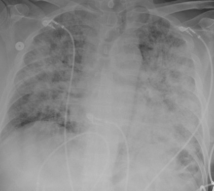
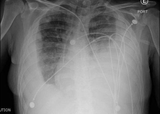
DOI: https://doi.org/10.21980/J8RW6NChest X-ray showed diffuse, patchy interstitial and alveolar infiltrates bilaterally concerning for Pneumocystis jirovecii(previously Pneumocystis carinii) pneumonia (PJP). The AP radiograph (top left figure) showed the classic “bat-wing” distribution on the left side. Repeat radiograph (bottom figure) one day after admission showed worsening of the infiltrates.
Suspicious Skin Lesion in an 11-Year-Old Male
DOI: https://doi.org/10.21980/J8JK9TThe patient had a 5 cm ulcerative lesion with raised borders and a yellow, “fatty” center. There was no active drainage, site tenderness, or lymphadenopathy.
Endocarditis
DOI: https://doi.org/10.21980/J8JP73Upright frontal radiograph of the chest demonstrated large pleural effusion on the left and moderate pleural effusion on the right as shown by the visible menisci on both sides (red arrows) with diffuse bilateral nodular densities (yellow dotted lines), consistent with septic pulmonary emboli. Computed tomography (CT) of the chest demonstrated multiple scattered lung nodules bilaterally containing internal foci of air cavitation (green dotted lines).
Fournier Gangrene
DOI: https://doi.org/10.21980/J89626The computed tomography (CT) of the abdomen and pelvis revealed significant subcutaneous gas tracking along the perineum and right gluteal region (orange outline) into the scrotum with associated scrotal edema (yellow arrow) and subcutaneous inflammatory fat stranding of 0.92 cm (red arrow) consistent with Fournier’s gangrene. There is early fluid loculation along the right medial gluteal cleft of 5.85 cm (green arrow) without a sizeable drainable abscess seen.
Sepsis Secondary to an Abdominal Wound Infection
DOI: https://doi.org/10.21980/J8PS60At completion of this case learners should be able to: 1) Recognize and differentiate between systemic inflammatory response syndrome, sepsis, severe sepsis, and septic shock. 2) Prepare an appropriate differential diagnosis for a patient with sepsis. 3) Demonstrate appropriate fluid resuscitation and antibiotic therapy for a septic patient. 4) Demonstrate appropriate vasopressor therapy for a septic patient. 5) Understand and apply the Surviving Sepsis Guidelines.
Necrotizing Soft Tissue Infection
DOI: https://doi.org/10.21980/J8X92TComputed tomography (CT) of the abdominal and pelvis with intravenous (IV) contrast revealed inflammatory changes, including gas and fluid collections within the ventral abdominal wall extending to the vulva, consistent with a necrotizing soft tissue infection.
A Case of Otomastoiditis
DOI: https://doi.org/10.21980/J8RK89The patient underwent computed tomography (CT) of the head which revealed opacification of the left middle ear (red arrow) and mastoid air cells (red circles). Additionally, there was thickening of the soft tissues of the external auditory canal (blue arrowhead), likely reflecting concurrent otitis externa. Based on the imaging, he was admitted for findings consistent with acute otomastoiditis.
Pediatric Pulmonary Abscess
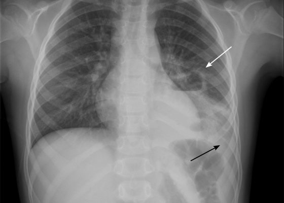
DOI: https://doi.org/10.21980/J83S6QUpright posterior-anterior plain chest films show a left lower lobe consolidation with an air-fluid level and a single septation consistent with a pulmonary abscess (white arrows). A small left pleural effusion was also present, seen as blunting of the left costophrenic angle and obscuration of the left hemidiaphragm (black arrows).


