• Registration
  • Login
JETem
  • Home
  • About
    • Aim and Scope
    • Our Team
    • Editorial Board
    • FAQ
  • Issues
    • Current Issue
    • Ahead of Print
    • Past Issues
  • Visual EM
    • Latest Visual EM
    • Search Visual EM
    • Thumbnail Library
  • For Authors
    • Instructions for Authors
    • Submit to JETem
    • Photo Consent
    • Policies
      • Peer Review Policy
      • Copyright Policy
      • Editorial Policy, Ethics and Responsibilities
      • Conflicts of Interest & Informed Consent
      • Open Access Policy
  • For Reviewers
    • Instructions for JETem Reviewers
    • Interested in Being a JETem Reviewer?
  • Topic
    • Abdominal / Gastroenterology
    • Administration
    • Board Review
    • Cardiology / Vascular
    • Clinical Informatics, Telehealth and Technology
    • Dermatology
    • EMS
    • Endocrine
    • ENT
    • Faculty Development
    • Genitourinary
    • Geriatrics
    • Hematology / Oncology
    • Infectious Disease
    • Miscellaneous
    • Neurology
    • Ob / Gyn
    • Ophthalmology
    • Orthopedics
    • Pediatrics
    • Pharmacology
    • Procedures
    • Psychiatry
    • Renal / Electrolytes
    • Respiratory
    • Social Determinants of Health
    • Toxicology
    • Trauma
    • Ultrasound
    • Urology
    • Wellness
    • Wilderness
  • Modality
    • Curricula
    • Innovations
    • Lectures
    • Oral Boards
      • Structured Interview
      • Communication Case
    • Podcasts
    • Simulation
    • Small Group Learning
    • Team Based Learning
    • Visual EM
  • Contact Us

Dermatology

Creative Commons images

Suspicious Skin Lesion in an 11-Year-Old Male

Rachel E Bonczek, MSN*, Kimberley M Farr, MD^ and Corrie E Chumpitazi, MD, MS‡

DOI: https://doi.org/10.21980/J8JK9TIssue 4:1 No ratings yet.
The patient had a 5 cm ulcerative lesion with raised borders and a yellow, “fatty” center. There was no active drainage, site tenderness, or lymphadenopathy.
Visual EMDermatologyInfectious Disease
Creative Commons images

Lightning Ground Current Injury: A Subtle Shocker

Gregory S Podolej, MD* and Naomi Nardi, MD*

DOI: https://doi.org/10.21980/J8KD1C Issue 4:1 No ratings yet.
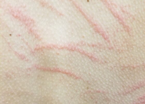
The first photograph demonstrates a dendritic blister (Lichtenburg figure) on the medial aspect of his right foot where the ground current injury entered the patient's foot. Although no data exists regarding the sensitivity or specificity of Lichtenberg figures as skin findings, they are considered pathognomonic for lightning injuries and are not produced by alternating current or industrial electrical injuries. The second photograph demonstrates a 4 x 3 cm area of petechiae where the ground current injury exited the patient.
WildernessDermatologyVisual EM
Creative Commons images

Pemphigoid Gestationis

Michael Niechayev, BS* and Lauren Sylwanowicz, MD*

DOI: https://doi.org/10.21980/J8MG9DIssue 3:4 No ratings yet.
Physical exam findings were significant for 1-3 cm diameter well-demarcated superficial ulcers on the patient’s abdomen and extremities, with mucosal sparing. Several small tense bullae were present on the bilateral inner thighs and numerous small reddish plaques were scattered over the patient’s back. Nikolsky’s sign was negative. No lymphadenopathy was noted.
DermatologyVisual EM
Creative Commons images

Hutchinson’s Sign

Lawrence Lau, MD*, Hannah Mirzakhani, BA^ and Nicholas Governatori, MD*

DOI: https://doi.org/10.21980/J8N040 Issue 3:1 No ratings yet.
The unilateral distribution of vesicular lesions over the patient's left naris, cheek, and upper lip are consistent with Herpes zoster reactivation with Hutchinson's sign.  Hutchinson's sign is a herpes zoster vesicle present on the tip or side of the nose.1 It reflects zoster involvement of the 1st branch of the trigeminal nerve, and is concerning for herpes zoster ophthalmicus.1 Herpes zoster vesicles may present as papular lesions or macular vesicles on an erythematous base.2,3 Emergent diagnosis must be made to prevent long-term visual sequelae.4
Infectious DiseaseDermatologyVisual EM
Creative Commons images

Erythema Migrans

Daniel Polvino, MD* and Grant Wei, MD*

DOI: https://doi.org/10.21980/J8QW7QIssue 2:4 No ratings yet.
History of present illness: A 28-year-old male presented to the emergency department with a chief complaint of two weeks of headache, chills, and numbness in his hands. He reported removing a tick from his upper back approximately two weeks ago, but did not know how long the tick had been embedded. His review of symptoms was otherwise unremarkable. Significant findings:
DermatologyInfectious DiseaseVisual EM

Chancre of Primary Syphilis

Adnan Riaz MD* and Grant Wei MD*

DOI: https://doi.org/10.21980/J83342 Issue 2:4 No ratings yet.
Physical examination revealed a non-tender, erythematous lesion on the glans penis, two similar adjacent satellite lesions, as well as tender inguinal lymphadenopathy. No penile discharge was noted.
Infectious DiseaseDermatologyGenitourinaryVisual EM
Creative Commons images

Steven-Johnson Syndrome

Pauline Joy F. Santos, MD* and Lauren Sylwanowicz, MD*

DOI: https://doi.org/10.21980/J8661W Issue 2:3 No ratings yet.
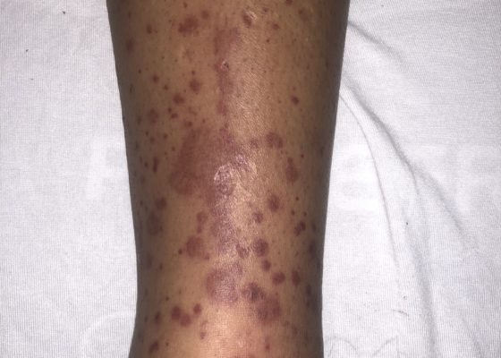
At presentation to the ED, a macular rash was notable on all four extremities, trunk and face, and involved mucous membranes of the oropharynx and vaginal introitus. The rash was painful, erythematous and purpuric with targetoid lesions. There were also multiple areas of sloughing and desquamation with a positive Nikolsky sign. Denudement totaled approximately 2% of total body surface area.
DermatologyVisual EM

Emergency Medicine Dermatology

Shannon Toohey, MD, MAEd*

DOI: https://doi.org/10.21980/J8DW21 Issue 2:2 No ratings yet.
By the end of this educational session, the learner will: 1) List the six primary types of rash (maculopapular, petechial/purpura, diffuse erythematous, non-erythematous, vesiculo-bullous, and pustular). 2) Be able to accurately describe various types of lesions and rashes with appropriate terminology; 3) Understand the use of the Modified Lynch Algorithm and how it can be used to narrow down the differential diagnosis in patients presenting with rash.
DermatologyTeam Based Learning (TBL)
Creative Commons images

Infectious Mononucleosis: Pharyngitis and Morbilliform Rash

Eric McCoy, MD, MPH* and Amal Shafi, BS*

DOI: https://doi.org/10.21980/J88C7H Issue 2:2 No ratings yet.
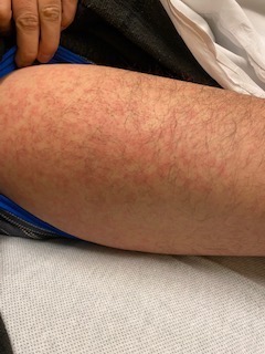
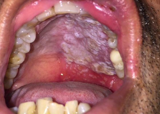
Her physical exam was significant for bilateral tonsillar exudates, cervical lymphadenopathy, and a morbilliform rash that included the palms (Figure 1-4). Laboratory testing was significant for white blood cell (WBC) count of 16.5 thous/mcl with an elevation in absolute lymphocytes of > 10 thous/mcl. The monospot and EBV (Epstein-Barr virus) panel were positive.
Infectious DiseaseDermatologyENTVisual EM
Creative Commons images

Herpes Zoster

Hamid Ehsani-Nia, BS, MS* and Robert Rowe, MD^

DOI: https://doi.org/10.21980/J8C301 Issue 2:2 No ratings yet.
The patient was in mild distress, afebrile, with stable vital signs. His physical exam revealed an erythematous, grouped vesicular rash in various stages of progression including erythematous papules, clear vesicles, and pustular vesicles. Few lesions were scabbed over. No signs of crusting or scarring were appreciated. The distribution encompassed the entire left T4 dermatome both posteriorly and anteriorly. No other rashes were appreciated elsewhere on the body.
Infectious DiseaseDermatologyVisual EM
‹2›
Page 2 of 3

JETem is an online, open access, peer-reviewed, journal-repository for EM educators. We are PMC Indexed.

Most Viewed

  • The Silent Saboteur: Teaching the Clinical Implications of Occult Hypoxemia & Social Determinants of Health via a Pulmonary Embolism Case
  • Diabetic Ketoacidosis and Necrotizing Soft Tissue Infection
  • My Broken Heart
  • Stabilization of Cardiogenic Shock for Critical Care Transport, a Simulation
  • Innovative Ultrasound-Guided Erector Spinae Plane Nerve Block Model for Training Emergency Medicine Physicians

Visit Our Collaborators

Creative Commons Licence
This work is licensed under a Creative Commons Attribution 4.0 International License.

About

Education

Learners should benefit from active learning. JETem accepts submissions of team-based learning, small group learning, simulation, podcasts, lectures, innovations, curricula, question sets, and visualEM.

Scholarship

We believe educators should advance through the scholarship of their educational work. JETem gives educators the opportunity to publish scholarly academic work so that it may be widely distributed, thereby increasing the significance of their results.

Links

  • Home
  • Aim and Scope
  • Current Issue
  • For Reviewers
  • Instructions for Authors
  • Contact Us

Newsletter

Sign up to receive updates from JETem regarding newly published issues and findings.

Copyright Creative Commons Attribution 4.0 International