MRI
Case Report of a Man with Right Eye Pain and Double Vision
DOI: https://doi.org/10.21980/J8KW7GABSTRACT: A 39-year-old previously healthy male presented with three days of right eye pressure and one day of binocular diplopia. He denied history of trauma, headache, or other neurological complaints. He had normal visual acuity, normal intraocular pressure, intact convergence, and no afferent pupillary defect. His neurologic examination was non-focal except for an inability to adduct the right eye past midline
A Boy with Rash and Joint Pain Diagnosed with Scurvy: A Case Report
DOI: https://doi.org/10.21980/J89H1XHis lower extremity magnetic resonance imaging (MRI) findings showed abnormal signals in his knees, which were most consistent with scurvy. The white arrows on the T1-weight sequence indicate hypointensity (decreased signal or darker region) of the knees. The white arrows in the T2-weighted short-tau inversion recovery (STIR) sequence indicate hyperintensity (increased signal or brighter region) in an MRI of the knees.
Erectile Dysfunction as a Presenting Symptom for Renal Cell Carcinoma
DOI: https://doi.org/10.21980/J8563BThe MRI showed extensive spondylotic changes suggestive of malignancy (red arrows) with severe spinal canal stenosis at the lumbar spine L3-L4 (purple arrows) level contributing to clumping of cauda equina nerve roots and severe bilateral neuroforaminal narrowing with diffuse disc bulges abutting the exiting nerve roots at multiple levels. Findings also showed a hypo-attenuated tumor (blue arrow) and hyper-attenuated loculated tumor (green arrow) consistent with renal cell carcinoma (RCC).
A Rare Cause of Pelvic Pain in a Teenage Girl
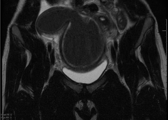
DOI: https://doi.org/10.21980/J87D0WDue to pain out of proportion to her exam, an ultrasound of her pelvis was obtained and showed a blood-filled distended uterus, or hematometrocolpos (white arrow), with a 4.9 cm right ovarian cyst (blue arrow). A pelvic magnetic resonance imaging (MRI) then revealed an obstructed right hemi-vagina, normal left uterus and vagina and ipsilateral renal agenesis (red arrow) with normal left kidney (double arrow) consistent with obstructed hemivagina, ipsilateral renal agenesis (OHVIRA) syndrome. The patient underwent surgical repair with complete resolution of symptoms.
Rare Rapidly Growing Thumb Lesion in a 12-Year-Old Male
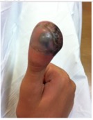
DOI: https://doi.org/10.21980/J8B92JHistory of present illness: A 12-year-old male presented to the emergency department with right thumb pain and a mass for four months (see images). He denied fevers, chills, change in appetite, or fatigue. He noted that the lesion was growing and “bleeds easily if bumped.” He denied any trauma to the thumb, except “hitting it” months ago while in football
‹2
Page 2 of 2


