• Registration
  • Login
JETem
  • Home
  • About
    • Aim and Scope
    • Our Team
    • Editorial Board
    • FAQ
  • Issues
    • Current Issue
    • Ahead of Print
    • Past Issues
  • Visual EM
    • Latest Visual EM
    • Search Visual EM
    • Thumbnail Library
  • For Authors
    • Instructions for Authors
    • Submit to JETem
    • Photo Consent
    • Policies
      • Peer Review Policy
      • Copyright Policy
      • Editorial Policy, Ethics and Responsibilities
      • Conflicts of Interest & Informed Consent
      • Open Access Policy
  • For Reviewers
    • Instructions for JETem Reviewers
    • Interested in Being a JETem Reviewer?
  • Topic
    • Abdominal / Gastroenterology
    • Administration
    • Board Review
    • Cardiology / Vascular
    • Clinical Informatics, Telehealth and Technology
    • Dermatology
    • EMS
    • Endocrine
    • ENT
    • Faculty Development
    • Genitourinary
    • Geriatrics
    • Hematology / Oncology
    • Infectious Disease
    • Miscellaneous
    • Neurology
    • Ob / Gyn
    • Ophthalmology
    • Orthopedics
    • Pediatrics
    • Pharmacology
    • Procedures
    • Psychiatry
    • Renal / Electrolytes
    • Respiratory
    • Social Determinants of Health
    • Toxicology
    • Trauma
    • Ultrasound
    • Urology
    • Wellness
    • Wilderness
  • Modality
    • Curricula
    • Innovations
    • Lectures
    • Oral Boards
      • Structured Interview
      • Communication Case
    • Podcasts
    • Simulation
    • Small Group Learning
    • Team Based Learning
    • Visual EM
  • Contact Us

Issue 8:2

How to Build a Low-Cost Video-Assisted Laryngoscopy Suite for Airway Management Training

Erin Falk, MD* and Adam Blumenberg, MD, MA*^

DOI: https://doi.org/10.21980/J8C068 Issue 8:2 No ratings yet.
Using an anatomically accurate airway simulator, by the end of a 20–30-minute instructional session, learners should be able to: 1) Understand proper positioning and use the video laryngoscope with dexterity, 2) identify airway landmarks via the video screen, and 3) demonstrate ability to intubate a simulated airway.
InnovationsProceduresRespiratory

Construction of Soft Prep Cadaver Pericardiocentesis Training Model and Implementation Among Emergency Medicine Residents

Kathryn Oskar, MD* and Dana Stearns, MD^

DOI: https://doi.org/10.21980/J87930 Issue 8:2 No ratings yet.
By the end of this session, residents will gain increased procedural competence and confidence with pericardiocentesis. Residents will be able to identify necessary supplies for the procedure, identify relevant surface anatomy and ultrasound views, and successfully aspirate fluid from model effusion.
Cardiology/VascularInnovationsProcedures

Flipping Tickborne Illnesses with Infographics

Daniel Johnson, DO* and Annahieta Kalantari, DO*

DOI: https://doi.org/10.21980/J83H12 Issue 8:2 No ratings yet.
After participation in this module, learners will be able to 1) list the causative agents for Lyme Disease, Babesiosis, Tularemia, Ehrlichiosis, Anaplasmosis, Tick Paralysis, Rocky Mountain Spotted Fever, and Powassan Virus, 2) identify different clinical features to distinguish the different presentations of tickborne illnesses, and 3) provide the appropriate treatments for each illness.
Infectious DiseaseSmall Group Learning

Peripartum Cardiomyopathy

Victoria L Morris, MD*, Carolina Mendoza, MD*, Gowri S Stevens, MD*, Jessica L Wilson, MD*and Adeola A Kosoko, MD*

DOI: https://doi.org/10.21980/J8ZS9M Issue 8:2 No ratings yet.
By the end of this simulation session, learners will be able to: 1) initiate a workup of a pregnant patient who presents with syncope, 2) accurately diagnose peripartum cardiomyopathy, 3) demonstrate care of a gravid patient in respiratory distress due to peripartum cardiomyopathy, 4) appropriately manage cardiogenic shock due to peripartum cardiomyopathy.
Cardiology/VascularOb/GynSimulation

Acute Exacerbation of COPD

Dominic Pappas, MD* and Amrita Vempati, MD*

DOI: https://doi.org/10.21980/J8V070 Issue 8:2 No ratings yet.
By the end of this simulation, learners will be able to (1) assess for causes of severe shortness of breath, (2) manage severe COPD exacerbation by administering appropriate medications, (3) identify worsening clinical status and initiate NIPPV, (4) assess the causes of hypoxia after establishing endotracheal intubation and, (5) identify indication for needle decompression and perform chest tube thoracostomy.
RespiratorySimulation

Botulism due to Drug Use

Timothy Hoffman, MD* and Jennifer Yee, DO*

DOI: https://doi.org/10.21980/J8Q93B Issue 8:2 No ratings yet.
ABSTRACT: Audience: This scenario was developed to educate emergency medicine residents on the diagnosis and management of wound botulism secondary to injection drug use.  Introduction: Botulism is a relatively rare cause of respiratory failure and descending weakness in the United States, caused by prevention of presynaptic acetylcholine release at the neuromuscular junction. This presentation has several mimics, including myasthenia gravis
SimulationToxicology
Creative Commons images

A Case Report of Subtle EKG Abnormalities in Acute Coronary Syndromes Indicative of Type One Myocardial Infarction

Paige Matijasich, BA*, Patrick Bruss, MD^, Gregory Reinhold, MD^ and Zachary Koppelmann, MD^

DOI: https://doi.org/10.21980/J8W06X Issue 8:2 No ratings yet.
The ECG does show multiple subtle abnormalities that in conjunction with his symptoms and risk factors are concerning for ischemia and/or occlusion of the coronary artery vessel. 1) ST depression in aVL. Although slight, the ST segment is below the TP segment or isoelectric point (blue circles). 2) Focal hyper QT waves. The T-waves in II, III, AVF V2, V3, and V4 are hyper acute, namely peaked and tall in relationship to the QRS. These are best displayed in leads II, III, and AVF where the T-waves are taller than the QRS amplitude (vertical blue lines). 3) Straightening off the ST segment. Multiple leads display a straight ST segment namely aVL, III, AVF, and V2 (red lines). Of note, the length of the straight ST segment is greater than 1/4 the amplitude of the QRS (purple lines). 4) Although subtle, these abnormalities are focal in nature.
Visual EMCardiology/Vascular
Creative Commons images

Case Report of Herpes Zoster Ophthalmicus with Concurrent Parotitis

Serena Tally, BS*, Michelle Brown, DO* and Edmund Hsu, MD*

DOI: https://doi.org/10.21980/J8R93N Issue 8:2 No ratings yet.
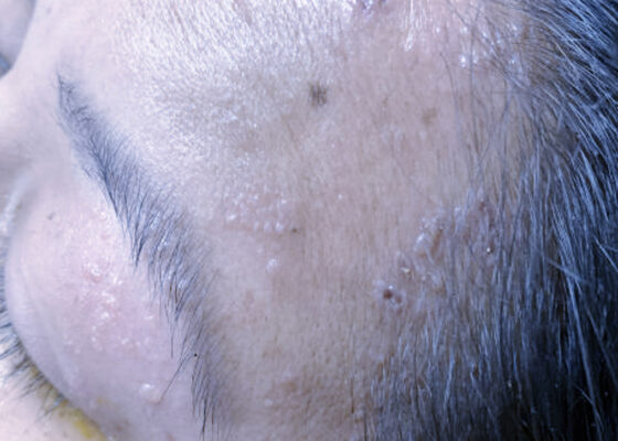
The presence of soft tissue stranding about the parotid gland suggested an underlying inflammatory or infectious process of the parotid gland. Cellulitis was considered as a possible diagnosis as well, given the presence of soft tissue stranding in the dermis that is adjacent to the parotid gland. Fortunately, no enhancement was seen in local muscles, fascia, or bones to suggest a myositis, fasciitis, or osteomyelitis. By using the anatomy of the patient and understanding the changes that occur on CT when inflammation is present, the appropriate depth and location of infection can be made, allowing for appropriate treatment regimens.
Visual EMInfectious DiseaseOphthalmology
Creative Commons images

A Man with Sore Throat—A Case Report

Nathan Mercado, BS*, Sawyer Schuljak, MD^, Daniel Ng, MD*^, Curtis Knight, MD*^, Allison Woodall, MD*^ and John Costumbrado, MD, MPH*^

DOI: https://doi.org/10.21980/J8MH0B Issue 8:2 No ratings yet.
Video laryngoscopy of the upper airway was performed two days after initial burn injury. The images obtained demonstrated laryngeal edema and inflammation near the epiglottis. The dot identifies the epiglottis and the asterix identifies the area of moderate thermal burns. Imaging also demonstrated adequate patency of airway and ruled out the need for intubation at that time.
Visual EMENTProcedures
Creative Commons images

The Continued Rise of Syphilis: A Case Report to Aid in Identification of the Great Imitator

Nicole Finney, MPH*, Eli Soyfer, MS*, Rory Schwan, MD* and Lindsey C Spiegelman, MD, MBA*

DOI: https://doi.org/10.21980/J8R93N Issue 8:2 No ratings yet.
Images taken of the bilateral palmar skin lesions at our institution showed multi-centimeter, well-demarcated, friable, verrucous, crusted plaques with overlying fine yellow crust. Lesions such as these are suspicious for syphilitic gummas seen with cutaneous tertiary syphilis.
Visual EMInfectious Disease

JETem is an online, open access, peer-reviewed, journal-repository for EM educators. We are PMC Indexed.

Most Viewed

  • The Silent Saboteur: Teaching the Clinical Implications of Occult Hypoxemia & Social Determinants of Health via a Pulmonary Embolism Case
  • Diabetic Ketoacidosis and Necrotizing Soft Tissue Infection
  • My Broken Heart
  • Stabilization of Cardiogenic Shock for Critical Care Transport, a Simulation
  • Innovative Ultrasound-Guided Erector Spinae Plane Nerve Block Model for Training Emergency Medicine Physicians

Visit Our Collaborators

Creative Commons Licence
This work is licensed under a Creative Commons Attribution 4.0 International License.

About

Education

Learners should benefit from active learning. JETem accepts submissions of team-based learning, small group learning, simulation, podcasts, lectures, innovations, curricula, question sets, and visualEM.

Scholarship

We believe educators should advance through the scholarship of their educational work. JETem gives educators the opportunity to publish scholarly academic work so that it may be widely distributed, thereby increasing the significance of their results.

Links

  • Home
  • Aim and Scope
  • Current Issue
  • For Reviewers
  • Instructions for Authors
  • Contact Us

Newsletter

Sign up to receive updates from JETem regarding newly published issues and findings.

Copyright Creative Commons Attribution 4.0 International