Issue 6:1
An Ultrasound-Guided Regional Anesthesia Elective for Emergency Medicine Residents
DOI: https://doi.org/10.21980/J8TP9BABSTRACT: Audience: This ultrasound-guided regional anesthesia elective is designed for emergency medicine residents. Length of Curriculum: The proposed length of this curriculum is over one week. Introduction: Ultrasound-guided regional anesthesia (UGRA) is a useful tool in the emergency department (ED) for managing painful conditions, and many programs have identified that these are useful skills for emergency providers; however, only about
Case Based Questions For Teaching EM Pharmacotherapy
DOI: https://doi.org/10.21980/J8PW61Our goals were to teach residents clinical applications of EM pharmacotherapy including drug selection and consideration of alternatives, interactions, and adverse effects, as well as to prepare them for pharmacotherapy questions on board examinations.
Design and Implementation of a Low-Cost Priapism Reduction Task Trainer
DOI: https://doi.org/10.21980/J8K64FBy the end of this educational session, learners should be able to 1) Verbalize the difference between low-flow and high-flow priapism 2) Describe the landmarks for a penile ring block and cavernosal aspiration/injection 3) Demonstrate the appropriate technique for performing a penile ring block, cavernosal aspiration, and cavernosal injection.
Botulism
DOI: https://doi.org/10.21980/J8FD0RBy the end of this simulation learners will be able to: 1) develop a differential for descending paralysis and recognize the signs and symptoms of botulism; 2) understand the importance of consulting public health authorities to obtain botulinum antitoxin in a timely fashion; 3) recognize that botulism will progress during the time period antitoxin is obtained. Early indications of respiratory compromise are expected to worsen during this time window.
Secondary learning objectives include: 4) employ advanced evaluation for neurogenic respiratory failure such as physical examination, negative inspiratory force (NIF), forced vital capacity (FVC), and partial pressure of carbon dioxide (pCO2), 5) discuss and review the pathophysiology of botulism, 6) discuss the epidemiology of botulism.
HIT-Heparin Induced Thrombocytopenia Simulation Case
DOI: https://doi.org/10.21980/J89Q0MAfter completing this simulated case, participants will be able to: 1) Obtain a detailed history that includes recent medications, medical, surgical, and social history to evaluate for HIT risk factors, 2) perform an adequate neurovascular exam including evaluation of motor function, sensation, skin color, pulses, and capillary refill, 3) order appropriate laboratory testing and imaging for diagnosis of thrombocytopenia and arterial occlusion, including bed side doppler or ultrasound, 4) discuss and recognize the symptoms of HIT and the contraindications of platelet and heparin administration in the emergency department, 5) avoid administration of heparin in the emergency department setting and recognize that platelets may worsen thrombus formation and lead to limb amputation, 6) select appropriate medications for treatment and determine appropriate disposition for a patient presenting with HIT, 7) demonstrate interpersonal communication with patient and family, 8) recognize that HIT with thrombosis is a potential complication in hospitalized patients and outpatient settings and is associated with high mortality rates.
Posterior Reversible Encephalopathy Syndrome (PRES)
DOI: https://doi.org/10.21980/J85W6CBy the end of the simulation, the learner will be able to: 1) manage an acute seizure 2) discuss imaging modalities to diagnose PRES 3) discuss medical management of PRES.
Approach to the Poisoned Patient
DOI: https://doi.org/10.21980/J8264SBy the end of the lecture, learners should be able to: 1) initiate the evaluation of a poisoned patient, 2) identify key interventions to support airway, breathing, and circulation, 3) identify the three components of risk assessment in the poisoned patient, 4) list the four options for gastric decontamination, and 5) select standard diagnostic labs and tests commonly used in evaluating poisoned patients.
Case Report of Thrombotic Thrombocytopenic Purpura in a Previously Healthy Adult
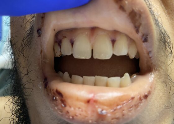
DOI: https://doi.org/10.21980/J8VK9MThe physical exam revealed globalized pallor of his skin as well as conjunctival pallor. Mucous membranes were found to be dry and pale with dried gingival hemorrhages apparent between teeth (image). Additionally, mild hepatosplenomegaly was noted. While in the ED, the patient’s urine was dark reddish-brown, which he then reported had been similarly discolored for the past 7 days.
A Case Report of Posterior Communicating Artery Aneurysm Presenting as Cranial Nerve 3 Palsy in a Young Female Patient with Migraines
DOI: https://doi.org/10.21980/J8QW83Physical exam revealed a right pupil that was dilated compared to the left pupil, though both pupils were reactive. The patient also had impaired medial gaze on the right and ptosis of the right eyelid. Exam was otherwise unremarkable.
Pulmonary Arteriovenous Malformation in a Patient with Suspected Hereditary Hemorrhagic Telangiectasia: A Case Report
DOI: https://doi.org/10.21980/J8M353Initial vital signs were unremarkable, including oxygen saturation of 98% on room air. The patient did not exhibit any signs of respiratory distress, and the lungs were clear to auscultation bilaterally. Labs were obtained, which showed normal hemoglobin at 15.8. Computed tomography (CT) of the chest (Video 1) showed a large left upper lobe arteriovenous malformation (AVM) with large feeding arteries and tortuous dilated draining veins (red arrow) measuring up to 3.8cm. Imaging also demonstrated nonspecific multifocal ground-glass opacities, which may have represented pulmonary hemorrhage (blue outline) from AVM without evidence of contrast extravasation to suggest active bleeding.
1›
Page 1 of 2


