CT
Elderly female with acute abdominal pain presenting with Superior Mesenteric Artery Thrombus
DOI: https://doi.org/10.21980/J82W52Computed tomography (CT) angiogram of the abdomen and pelvis revealed a superior mesenteric artery (SMA) thrombosis 5 cm from the origin off of the abdominal aorta. As seen in the sagittal view, there does not appear to be any contrast 5 cm past the origin of the SMA. On the axial views, you can trace the SMA until the point that there is no longer any contrast visible which indicates the start of the thrombus. The SMA does not appear to be reconstituted. There was normal flow to the celiac artery. (See annotated images).
Viridans streptococci Intracranial Abscess Masquerading as Metastatic Disease
DOI: https://doi.org/10.21980/J8CH05A non-contrast CT (Figure 1) revealed a large hypoattenuating left parietal lesion. When the CT was enhanced with intravenous contrast (Figure 2), the same lesion showed peripheral rim enhancement, suggestive of a brain abscess.
Pneumomediastinum After Cervical Stab Wound
DOI: https://doi.org/10.21980/J87P79Anteroposterior (AP) chest X-ray showed subcutaneous emphysema of the neck, surrounding the trachea (red arrows), right side greater than left, and a streak of gas adjacent to the aortic arch (white arrow). Computed tomography angiogram (CTA) of the neck showed air outside of the trachea, positive for pneumomediastinum (blue arrows).
Computed Tomography and Ultrasound Diagnosis of Spontaneous Subcapsular Renal Hematoma
DOI: https://doi.org/10.21980/J8062DBedside ultrasound was performed and demonstrated a hypoechoic area within the left kidney (images not shown). The non-contrast computed tomography (CT) of the abdomen and pelvis shows a significantly enlarged left kidney and a region of high-attenuation encapsulating the left kidney, concerning for acute hemorrhage.
Perianal Abscess
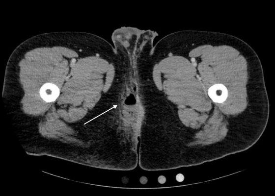
DOI: https://doi.org/10.21980/J8QP81Computed Tomography (CT) of the Pelvis with intravenous (IV) contrast revealed a 5.7 cm x 2.4 cm air-fluid collection in the right perianal soft tissue along the right gluteal cleft, with surrounding fat stranding, consistent with a perianal abscess with cellulitis.
Acute Pyelonephritis with Perinephric Stranding on CT
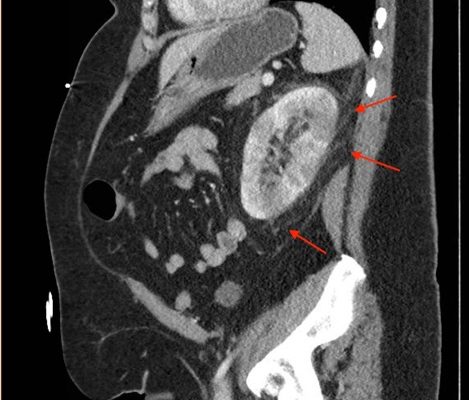
DOI: https://doi.org/10.21980/J8BH0VA CT abdomen and pelvis with IV contrast showed neither nephrolithiasis nor diverticulitis, and instead showed heterogeneous enhancement of the left kidney with mild edematous enlargement and striated left nephrogram. Significant perinephric stranding (red arrows) was also noted and was consistent with severe acute pyelonephritis.
Empyema
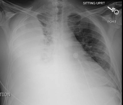
DOI: https://doi.org/10.21980/J86P9RThe chest X-ray shows a large fluid collection in the right lung demonstrated by the opacification that blunts the costophrenic angle on the right side. There is also a meniscus present, which is generally indicative of fluid. Chest computed tomography (CT) demonstrated an infiltrate with a mixture of densities within the same collection, consistent with a loculated effusion and concerning for an empyema.
Oropharynx Ulceration
DOI: https://doi.org/10.21980/J87W60The photograph demonstrates an area of ulcerative tissue at the left palatine tonsil without surrounding erythema or purulent drainage. The computed tomography (CT) scan shows a large ulceration of the left soft palate and palatine tonsil (red arrow). There is no evidence of skull base osteomyelitis. There is suppurative lymphadenopathy with partial left jugular vein compression due to mass effect (yellow highlight). There is mild nasopharyngeal airway narrowing with architectural distortion (blue arrow), but no other evidence of airway obstruction.


