• Registration
  • Login
JETem
  • Home
  • About
    • Aim and Scope
    • Our Team
    • Editorial Board
    • FAQ
  • Issues
    • Current Issue
    • Ahead of Print
    • Past Issues
  • Visual EM
    • Latest Visual EM
    • Search Visual EM
    • Thumbnail Library
  • For Authors
    • Instructions for Authors
    • Submit to JETem
    • Photo Consent
    • Policies
      • Peer Review Policy
      • Copyright Policy
      • Editorial Policy, Ethics and Responsibilities
      • Conflicts of Interest & Informed Consent
      • Open Access Policy
  • For Reviewers
    • Instructions for JETem Reviewers
    • Interested in Being a JETem Reviewer?
  • Topic
    • Abdominal / Gastroenterology
    • Administration
    • Board Review
    • Cardiology / Vascular
    • Clinical Informatics, Telehealth and Technology
    • Dermatology
    • EMS
    • Endocrine
    • ENT
    • Faculty Development
    • Genitourinary
    • Geriatrics
    • Hematology / Oncology
    • Infectious Disease
    • Miscellaneous
    • Neurology
    • Ob / Gyn
    • Ophthalmology
    • Orthopedics
    • Pediatrics
    • Pharmacology
    • Procedures
    • Psychiatry
    • Renal / Electrolytes
    • Respiratory
    • Social Determinants of Health
    • Toxicology
    • Trauma
    • Ultrasound
    • Urology
    • Wellness
    • Wilderness
  • Modality
    • Curricula
    • Innovations
    • Lectures
    • Oral Boards
      • Structured Interview
      • Communication Case
    • Podcasts
    • Simulation
    • Small Group Learning
    • Team Based Learning
    • Visual EM
  • Contact Us

CT

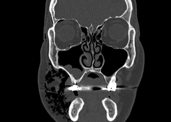
Dental Free Air CT Coronal Unannotated. JETem 2025

A Case Report of Facial Swelling and Crepitus Following a Dental Procedure

Shady Mikhail, MD*, George Mina, MD^, Alisa Wray MD, MAEd† and Danielle Matonis, MD†

DOI: https://doi.org/10.21980/J83W8H Issue 10:3 No ratings yet.
Given the physical exam findings of crepitus on the right neck up to the right lower eyelid, a maxillofacial CT scan without contrast was performed. It revealed diffuse subcutaneous air within the soft tissues of the face and neck and free air within the pre-septal soft tissue of the right eye, appearing as hyperlucent (dark) areas on CT within the soft tissue planes (blue outline). It showed no evidence of post-septal free air. A single-view chest X-ray was also performed and was unremarkable except for incompletely imaged soft tissue gas in the right lower neck (blue outline). On flexible fiberoptic laryngoscopy performed by ENT, the oropharynx appeared diffusely edematous and narrowed.
Visual EMCurrent IssueENT
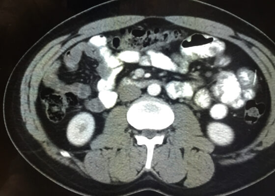
Iatrogenic Bowel Perforation Following Dental Procedure. CT. Unannotated. JETem 2025

Case Report: Iatrogenic Bowel Perforation Following Dental Procedure

Claire DeLong, DO* and Frederick Fiesseler, DO*

DOI: https://doi.org/10.21980/J8CD38 Issue 10:3 No ratings yet.
The patient’s abdominal CT demonstrated a metallic foreign body in the left side of the abdomen within the small bowel, without surrounding induration or abscess. Radiology questioned whether the metallic foreign object perforated the bowel. Seen in the cross-sectional CT image, there is a hyperdense linear structure transversing the small intestinal wall, given that a portion of the structure was located outside of the lumen of the bowel.
Visual EMAbdominal/GastroenterologyCurrent Issue
CSpine Fracture CT Axial Unannotated. JETem 2025

A Case Report of an Unstable C-spine Fracture in the Emergency Department

Jinho Jung, BS*, Tyler Rigdon, MD^, Alisa Wray, MD, MAEd^ and Danielle Matonis, MD^

DOI: https://doi.org/10.21980/J8SK90 Issue 10:2 No ratings yet.
The initial workup in the ED showed an acute displaced fracture of the left occipital condyle (CT-coronal, fracture of the left occipital condyle, red arrow; displacement, orange line), a shattered left lateral mass with involvement of the vertebral canal (CT-axial, red arrow), and malalignment of the craniocervical junction (CT-sagittal, red outline). The CT angiogram head and neck showed a possible irregularity in the left vertebral artery. The CT head without contrast had no significant findings.
Visual EM
Hydropic Gallbladder. CT Coronal Unannotatd. JETem 2025

A Case Report of Hydropic Gallbladder Presenting as Right Lower Quadrant Abdominal Pain

Savannah Tan, MD*, Zoe Adams, BA^, Scott Rudkin, MD, MBA* and Danielle Matonis, MD*

DOI: https://doi.org/10.21980/J8DD26 Issue 10:2 No ratings yet.
Computed tomography (CT) of the abdomen and pelvis with contrast was ordered, and general surgery was consulted for the initial working diagnosis of acute appendicitis. However, the CT scan resulted with findings of a markedly distended gallbladder measuring approximately 14.5 x 4 centimeters (cm) with marked gallbladder wall thickening (magenta) and pericholecystic fat stranding (cyan). The appendix was not dilated and had no inflammatory changes or edema. Follow-up right upper quadrant ultrasound confirmed the diagnosis of acute cholecystitis.
Visual EMAbdominal/GastroenterologyInfectious Disease
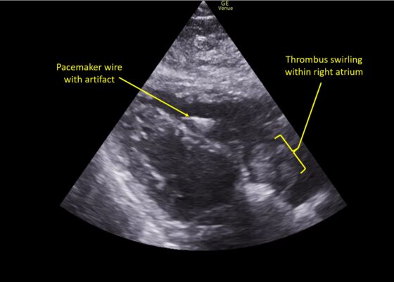
Wolff A et al. Right atrial thrombosis. Parasternal long US 3 annotated

A Case Report of Right Atrial Thrombosis Complicated by Multiple Pulmonary Emboli: POCUS For the Win!

Andrea Wolff, MD*, Evan Leibner, MD* and Jill Gualdoni, MD^

DOI: https://doi.org/10.21980/J8TM07 Issue 10:1 No ratings yet.
Pulmonary POCUS was performed by the ED physician (GE Venue, C1-5-RS 5MHz curvilinear transducer), and lung examination was unremarkable with no pleural effusion, pneumothorax, or infiltrate. Subxiphoid views (GE Venue, 3Sc-RS 4MHz phased-array transducer) were obtained because this patient’s COPD with severe pulmonary hyperexpansion made parasternal and apical 4-chamber views suboptimal. A large thrombus can be seen within the right atrium (movie 1, images 1, 2). This has a serpiginous, rounded appearance and is mobile, appearing to swirl within the right atrium with intermittent extrusion through the tricuspid valve. A pacemaker wire is also visible within the right ventricle as a non-moving, hyperechoic, linear structure with posterior enhancement artifact. Pericardial effusion is not present.
Visual EMCardiology/VascularUltrasound
RPA Neck. CT Lateral. Unannotated. JETem 2025

Retropharyngeal Abscess in an Adult Patient Presenting with Neck Fullness and Dysphagia: A Case Report

Justin Rederer, DO*, Tanner Folster, DO^ and Sara Dimeo, MD, MEHP^

DOI: https://doi.org/10.21980/J8M36G Issue 10:1 No ratings yet.
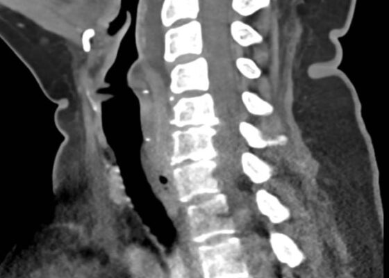
Contrast-enhanced CT soft tissue of the neck showed evidence of a prevertebral/retropharyngeal fluid collection, extending from the odontoid tip to the inferior C4 vertebral body margin, measuring 5.4 x 1.0 x 3.3 centimeters (cm) in size (yellow lines) without gross airway narrowing.
Visual EMENT
Chest wall abscess. Photograph unannotated. JETem 2024

A Man With Chest Pain After An Assault – A Case Report

Mi Song Kim, MD*, Francis Gan, MD*, Karl Nimtz, MD*, Daniel Ng, MD*^ and John Costumbrado, MD, MPH*^

DOI: https://doi.org/10.21980/J8J93S Issue 9:3 No ratings yet.
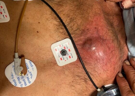
On exam, we found a suspected chest wall abscess with surrounding erythema (blue arrow). The patient underwent CT of the chest which showed a comminuted displaced midsternal fracture (yellow arrow) with moderate fluid and air anteriorly (red arrow), consistent with an abscess. His laboratory results had no significant abnormalities.
Visual EMInfectious DiseaseTrauma
Gastric Emphysema. Coronal CT annotated. JETem 2024

A Case Report Evaluating Gastric Emphysema versus Emphysematous Gastritis

Anna Nguyen*, Mark Slader, MD ^, Lindsey Spiegelman, MD, MBA^

DOI: https://doi.org/10.21980/J8ZH26Issue 9:2 No ratings yet.
A CT scan of the abdomen and pelvis was obtained and revealed gas within the gastric wall at the fundus (blue arrows), concerning for gastric emphysema versus emphysematous gastritis. There was no gastric wall thickening, free air, bowel obstruction, drainable fluid collection, or evidence of portal venous gas. Incidentally, hepatomegaly and likely hepatic steatosis were also noted.
Visual EM
Adult Intussusception. CT Axial Unannotated. JETem 2024

Telescoping into Adulthood: A Case Report of Intussusception in an Adult Patient

Neena Joy, DO*, and Laura Kolster, DO*

DOI: https://doi.org/10.21980/J8Q06C Issue 9:2 No ratings yet.
Computed tomography imaging of the abdomen and pelvis with intravenous and oral contrasts was obtained. In the axial view, one will see a concentric ring formed by layers of bowel, mesenteric vessels, and fat (red arrow and circle); this is the equivalent of the ultrasonographic “target sign.” The inner ring (blue arrow) represents the lead point causing telescoping of the bowel. One can see that the proximal bowel is dilated (yellow arrow). In the coronal view, one can see an obstructive mass, also known as the lead point (red arrow), located in the lumen of the descending colon. Located proximal to the lead point are dilated loops of bowel with edematous changes and fat stranding (pink circle). The proximal portion of the bowel will take on a concentric appearance with the telescoping loop of bowel.
Abdominal/GastroenterologyVisual EM
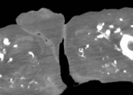
R Retroperitoneal Hematoma US 1 Unannotated. JETem 2023

Trauma by Couch: A Case Report of a Massive Traumatic Retroperitoneal Hematoma

Cassandra Smith, BSN *, Graham Stephenson, MD*, Alisa Wray, MD, MAEd* and Matthew Hatter, BS*

DOI: https://doi.org/10.21980/J84D2QIssue 8:3 No ratings yet.
Upon arrival at the trauma center, a FAST revealed a large, well-circumscribed abnormality (red outline) deep to the liver (blue outline and star) and gallbladder (green outline and star). The right kidney and hepatorenal space were not clearly visualized. The remainder of the FAST showed no free fluid in the splenorenal space, pelvis, and no pericardial effusion. He had lung sliding bilaterally.
TraumaVisual EM
1›»
Page 1 of 13

JETem is an online, open access, peer-reviewed, journal-repository for EM educators. We are PMC Indexed.

Most Viewed

  • A Longitudinal, Practical Curriculum for Faculty Development as New Coaches in Graduate Medical Education
  • Cognitive Errors and Debiasing
  • Critical Care Transport: Blunt Polytrauma in Pregnancy
  • Beta-Blocker Toxicity
  • A Comprehensive and Modality Diverse Cervical Spine and Back Musculoskeletal Physical Exam Curriculum for Medical Students

Visit Our Collaborators

Creative Commons Licence
This work is licensed under a Creative Commons Attribution 4.0 International License.

About

Education

Learners should benefit from active learning. JETem accepts submissions of team-based learning, small group learning, simulation, podcasts, lectures, innovations, curricula, question sets, and visualEM.

Scholarship

We believe educators should advance through the scholarship of their educational work. JETem gives educators the opportunity to publish scholarly academic work so that it may be widely distributed, thereby increasing the significance of their results.

Links

  • Home
  • Aim and Scope
  • Current Issue
  • For Reviewers
  • Instructions for Authors
  • Contact Us

Newsletter

Sign up to receive updates from JETem regarding newly published issues and findings.

Copyright Creative Commons Attribution 4.0 International