• Registration
  • Login
JETem
  • Home
  • About
    • Aim and Scope
    • Our Team
    • Editorial Board
    • FAQ
  • Issues
    • Current Issue
    • Ahead of Print
    • Past Issues
  • Visual EM
    • Latest Visual EM
    • Search Visual EM
    • Thumbnail Library
  • For Authors
    • Instructions for Authors
    • Submit to JETem
    • Photo Consent
    • Policies
      • Peer Review Policy
      • Copyright Policy
      • Editorial Policy, Ethics and Responsibilities
      • Conflicts of Interest & Informed Consent
      • Open Access Policy
  • For Reviewers
    • Instructions for JETem Reviewers
    • Interested in Being a JETem Reviewer?
  • Topic
    • Abdominal / Gastroenterology
    • Administration
    • Board Review
    • Cardiology / Vascular
    • Clinical Informatics, Telehealth and Technology
    • Dermatology
    • EMS
    • Endocrine
    • ENT
    • Faculty Development
    • Genitourinary
    • Geriatrics
    • Hematology / Oncology
    • Infectious Disease
    • Miscellaneous
    • Neurology
    • Ob / Gyn
    • Ophthalmology
    • Orthopedics
    • Pediatrics
    • Pharmacology
    • Procedures
    • Psychiatry
    • Renal / Electrolytes
    • Respiratory
    • Social Determinants of Health
    • Toxicology
    • Trauma
    • Ultrasound
    • Urology
    • Wellness
    • Wilderness
  • Modality
    • Curricula
    • Innovations
    • Lectures
    • Oral Boards
      • Structured Interview
      • Communication Case
    • Podcasts
    • Simulation
    • Small Group Learning
    • Team Based Learning
    • Visual EM
  • Contact Us

Latest Articles

simulation icon

High-Fidelity Simulation with Transvaginal Ultrasound in the Emergency Department

Levi Filler, DO* and Katrina Lettang, MD*

DOI: https://doi.org/10.21980/J8606Q Issue 9:3 No ratings yet.
By the end of the session, learners should be able to 1) recognize the clinical indications for transvaginal ultrasound in the ED, 2) practice the insertion, orientation, and sweeping motions used to perform a TVPOCUS study, 3) interpret transvaginal ultrasound images showing an IUP or alternative pathologies, and 4) understand proper barrier, disinfection, and storage techniques for endocavitary probes.
Ob/GynProceduresSimulation
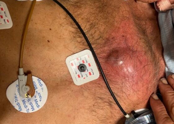
Chest wall abscess. Photograph unannotated. JETem 2024
visualem icon

A Man With Chest Pain After An Assault – A Case Report

Mi Song Kim, MD*, Francis Gan, MD*, Karl Nimtz, MD*, Daniel Ng, MD*^ and John Costumbrado, MD, MPH*^

DOI: https://doi.org/10.21980/J8J93S Issue 9:3 No ratings yet.
On exam, we found a suspected chest wall abscess with surrounding erythema (blue arrow). The patient underwent CT of the chest which showed a comminuted displaced midsternal fracture (yellow arrow) with moderate fluid and air anteriorly (red arrow), consistent with an abscess. His laboratory results had no significant abnormalities.
Visual EMInfectious DiseaseTrauma
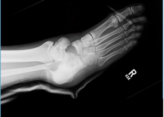
Subtalar Dislocation. sp manipulation unannotated. JETem 2024
visualem icon

A Case Report of Lateral Subtalar Dislocation: Emergency Medicine Assessment, Management and Disposition

Alexander Maybury, MD* and Taylor Isenberg, DO*

DOI: https://doi.org/10.21980/J8SS8P Issue 9:3 No ratings yet.
In a lateral subtalar dislocation, the navicular bone (red bone in 3D anatomy image) and the calcaneus (yellow bone in 3D anatomy image) dislocate laterally in relation to the talus (lavender bone in 3D anatomy image). Plain film oblique and lateral X-rays demonstrate the initial dislocation (talus in red, navicular in blue). It is clear in the initial lateral view that there is loss of the talar/navicular articulation (noted by red arrow). The anterior-posterior x-ray is more challenging to discern the anatomy; however, the talus (red dot) is laterally displaced in comparison to the navicular (blue dot).
Visual EMOrthopedicsProcedures
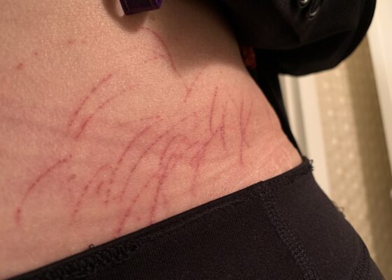
Dematographia
visualem icon

A Case Report of Dermatographia

Mahika Patlola*, Aanchal A Shah, BA^, Thor S. Stead, ScB† and Latha Ganti, MD, MS, MBA† **

DOI: https://doi.org/10.21980/J8P05P Issue 9:3 No ratings yet.
Physical examination was unremarkable except for the urticaria on the right aside of her abdomen (white arrow) with overlying excoriations (stars). Of note, there were no burrows, papules or vesicles in the typical locations including the webs of the fingers, wrists, axillae, areolae, or genitalia. Examination of the linear dermatographia clearly revealed superficial wheals, versus underlying serpiginous lesions.
Visual EMDermatology
innovations icon

Modification of an Airway Training Mannequin to Teach Engagement of the Hyoepiglottic Ligament

Richard Tumminello, DO* and Daniel Patino-Calle, MD*

DOI: https://doi.org/10.21980/J8R06P Issue 9:2 No ratings yet.
By the end of this education session, participants should be able to: 1) identify relevant airway anatomy during intubation, including base of the tongue, epiglottis, midline vallecular fold, anterior arytenoids; 2) appreciate the value of a stepwise anatomically guided approach to intubation; 3) become familiar with the midline vallecular fold and underlying anatomy, including the hyoepiglottic ligament, and how proper placement of the laryngoscope can result in improved glottic visualization.
InnovationsProceduresRespiratory
innovations icon

A Realistic, Low-Cost Simulated Automated Chest Compression Device

Jessica Joyce, BS*, Elyse Fults, MD^, Julia Rajan, BS†, Alexandra Plezia, MA**,  Carolyn Clayton, MD^ and Sara M Hock, MD^

DOI: https://doi.org/10.21980/J8M63C Issue 9:2 No ratings yet.
By the end of this educational session using a resuscitation trainer or high-fidelity manikin, learners should be able to: 1) recognize appropriate application of simulated ACCD to an ongoing resuscitation case; 2) demonstrate proper positioning of simulated ACCD in manikin model and 3) integrate simulated ACCD to provide compressions appropriately throughout cardiac arrest scenario.
Cardiology/VascularInnovationsProcedures
Show More

JETem is an online, open access, peer-reviewed, journal-repository for EM educators. We are PMC Indexed.

Most Viewed

  • A Longitudinal, Practical Curriculum for Faculty Development as New Coaches in Graduate Medical Education
  • Cognitive Errors and Debiasing
  • Critical Care Transport: Blunt Polytrauma in Pregnancy
  • Beta-Blocker Toxicity
  • A Comprehensive and Modality Diverse Cervical Spine and Back Musculoskeletal Physical Exam Curriculum for Medical Students

Visit Our Collaborators

Creative Commons Licence
This work is licensed under a Creative Commons Attribution 4.0 International License.

About

Education

Learners should benefit from active learning. JETem accepts submissions of team-based learning, small group learning, simulation, podcasts, lectures, innovations, curricula, question sets, and visualEM.

Scholarship

We believe educators should advance through the scholarship of their educational work. JETem gives educators the opportunity to publish scholarly academic work so that it may be widely distributed, thereby increasing the significance of their results.

Links

  • Home
  • Aim and Scope
  • Current Issue
  • For Reviewers
  • Instructions for Authors
  • Contact Us

Newsletter

Sign up to receive updates from JETem regarding newly published issues and findings.

Copyright Creative Commons Attribution 4.0 International