• Registration
  • Login
JETem
  • Home
  • About
    • Aim and Scope
    • Our Team
    • Editorial Board
    • FAQ
  • Issues
    • Current Issue
    • Ahead of Print
    • Past Issues
  • Visual EM
    • Latest Visual EM
    • Search Visual EM
    • Thumbnail Library
  • For Authors
    • Instructions for Authors
    • Submit to JETem
    • Photo Consent
    • Policies
      • Peer Review Policy
      • Copyright Policy
      • Editorial Policy, Ethics and Responsibilities
      • Conflicts of Interest & Informed Consent
      • Open Access Policy
  • For Reviewers
    • Instructions for JETem Reviewers
    • Interested in Being a JETem Reviewer?
  • Topic
    • Abdominal / Gastroenterology
    • Administration
    • Board Review
    • Cardiology / Vascular
    • Clinical Informatics, Telehealth and Technology
    • Dermatology
    • EMS
    • Endocrine
    • ENT
    • Faculty Development
    • Genitourinary
    • Geriatrics
    • Hematology / Oncology
    • Infectious Disease
    • Miscellaneous
    • Neurology
    • Ob / Gyn
    • Ophthalmology
    • Orthopedics
    • Pediatrics
    • Pharmacology
    • Procedures
    • Psychiatry
    • Renal / Electrolytes
    • Respiratory
    • Social Determinants of Health
    • Toxicology
    • Trauma
    • Ultrasound
    • Urology
    • Wellness
    • Wilderness
  • Modality
    • Curricula
    • Innovations
    • Lectures
    • Oral Boards
      • Structured Interview
      • Communication Case
    • Podcasts
    • Simulation
    • Small Group Learning
    • Team Based Learning
    • Visual EM
  • Contact Us

Latest Articles

Creative Commons images
visualem icon

Woman Swallows a “Handful of Pills”

Sarah E Mott, MD*, Michael Paddock, DO, MS*^ and Jessie Nelson, MD*^

DOI: https://doi.org/10.21980/J8V64XIssue 3:3 No ratings yet.
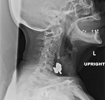
Soft tissue lateral X-ray of neck was performed. The lateral soft tissue X-ray of the neck showed a metallic foreign body at the level cricoid.
Abdominal/GastroenterologyVisual EM
Creative Commons images
visualem icon

Lisfranc Injury

Lisa Schwartz, BS*, Nelly Tan, MD^ and John Costumbrado, MD, MPH*

DOI: https://doi.org/10.21980/J8QD1MIssue 3:3 No ratings yet.
The frontal view of the right foot showed divergent dislocation of the second through fifth metatarsal bones (red outlines) consistent with Lisfranc injury. Though the Lisfranc ligament is not visualized by radiograph, the yellow markings represent the location of the Lisfranc ligament between the medial cuneiform (blue dot) and the base of the second metatarsal bone.  The first metatarsal and the medial cuneiform remain congruent. The lateral view shows dorsal dislocation of the midfoot (pink circle) consistent with instability. There is associated extensive midfoot soft tissue swelling.
OrthopedicsVisual EM
Creative Commons images
visualem icon

Incidental Hiatal Hernia on Chest X-ray

Ahmed Farhat, BS* and Daryn Towle, MD*

DOI: https://doi.org/10.21980/J8KP8SIssue 3:3 No ratings yet.
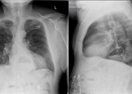
The two-view chest X-ray shows mild opacification of the bilateral lower lobes concerning for pneumonia (red arrows). Incidental retrocardiac opacity with air-fluid level consistent with large hiatal hernia is also observed (green arrow).
Abdominal/GastroenterologyVisual EM
Creative Commons images
visualem icon

Button Battery in Esophagus

Sha Yan, DO* and Ernest Leva, MD*

DOI: https://doi.org/10.21980/J8FW6VIssue 3:3 No ratings yet.
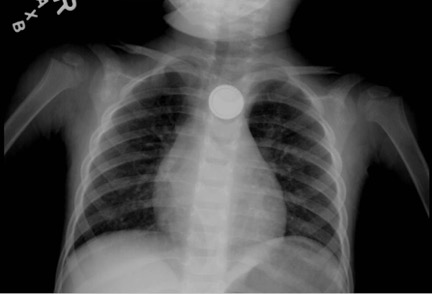
Chest radiograph showed the presence of a round radiopaque foreign body in the mid-chest. It was suspected to be in the esophagus rather than in the trachea due to the en-face positioning of the foreign body. The foreign body demonstrated two concentric ring circles concerning for a “double ring” or “halo" sign, which was suggestive of the presence of a button battery rather than a coin.
Abdominal/GastroenterologyVisual EM
Creative Commons images
visualem icon

Pediatric Foreign Body Aspiration

Sha Yan, DO* and Uchechi Azubuine, MD^

DOI: https://doi.org/10.21980/J8B648Issue 3:3 No ratings yet.
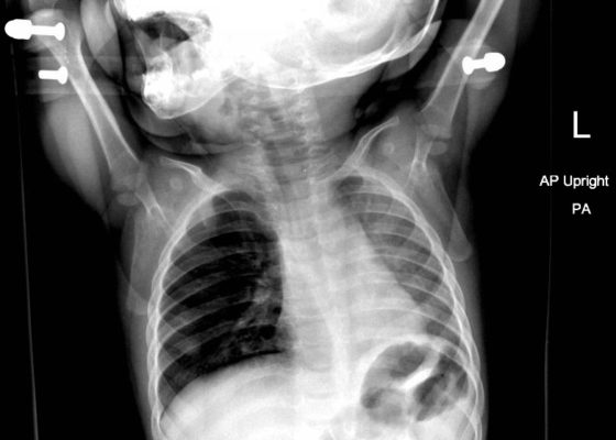
Chest radiograph showed increased radiolucency (red arrow) and flattening of the diaphragm on the right side (blue arrow) consistent with hyperinflation of the right lung, as well as left mediastinal shift (green arrow), indicating obstruction.
PediatricsRespiratoryVisual EM
Creative Commons images
visualem icon

Achalasia: An Uncommon Presentation with Classic Imaging

Joseph Adamson, BS*, Mina Altwail, MD^ and Shanna Jones, MD^

DOI: https://doi.org/10.21980/J86D2BIssue 3:3 No ratings yet.
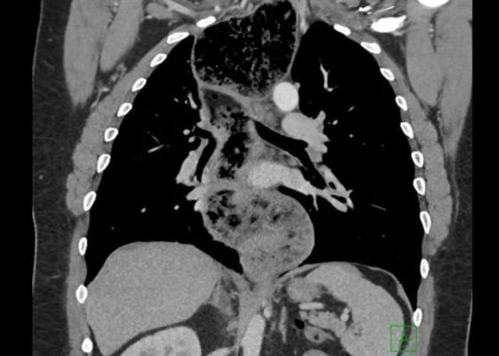
The chest X-ray demonstrated a markedly widened mediastinum (red brackets), raising concern for thoracic aortic aneurysm/aortic dissection, which prompted labs and contrast-enhanced computed tomography (CT) of the chest. The CT revealed a dilated proximal esophagus that narrowed distally (yellow tracing and red arrow), with particulate material, mass-effect on the trachea (purple outline), and bilateral patchy opacities suggesting aspiration. Barium esophagram showed a drastically dilated esophagus filled with contrast (yellow arrow), terminating into the classic “bird’s beak sign” (red arrow) at the lower esophageal sphincter (LES). Esophageal manometry later confirmed achalasia, proving that widened mediastina can have unexpected etiologies.
Abdominal/GastroenterologyVisual EM
Show More

JETem is an online, open access, peer-reviewed, journal-repository for EM educators. We are PMC Indexed.

Most Viewed

  • A Longitudinal, Practical Curriculum for Faculty Development as New Coaches in Graduate Medical Education
  • Cognitive Errors and Debiasing
  • Critical Care Transport: Blunt Polytrauma in Pregnancy
  • Beta-Blocker Toxicity
  • A Comprehensive and Modality Diverse Cervical Spine and Back Musculoskeletal Physical Exam Curriculum for Medical Students

Visit Our Collaborators

Creative Commons Licence
This work is licensed under a Creative Commons Attribution 4.0 International License.

About

Education

Learners should benefit from active learning. JETem accepts submissions of team-based learning, small group learning, simulation, podcasts, lectures, innovations, curricula, question sets, and visualEM.

Scholarship

We believe educators should advance through the scholarship of their educational work. JETem gives educators the opportunity to publish scholarly academic work so that it may be widely distributed, thereby increasing the significance of their results.

Links

  • Home
  • Aim and Scope
  • Current Issue
  • For Reviewers
  • Instructions for Authors
  • Contact Us

Newsletter

Sign up to receive updates from JETem regarding newly published issues and findings.

Copyright Creative Commons Attribution 4.0 International