Latest Articles
Posterior Reversible Encephalopathy Syndrome (PRES)
DOI: https://doi.org/10.21980/J85W6CBy the end of the simulation, the learner will be able to: 1) manage an acute seizure 2) discuss imaging modalities to diagnose PRES 3) discuss medical management of PRES.
Approach to the Poisoned Patient
DOI: https://doi.org/10.21980/J8264SBy the end of the lecture, learners should be able to: 1) initiate the evaluation of a poisoned patient, 2) identify key interventions to support airway, breathing, and circulation, 3) identify the three components of risk assessment in the poisoned patient, 4) list the four options for gastric decontamination, and 5) select standard diagnostic labs and tests commonly used in evaluating poisoned patients.
Case Report of Thrombotic Thrombocytopenic Purpura in a Previously Healthy Adult
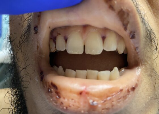
DOI: https://doi.org/10.21980/J8VK9MThe physical exam revealed globalized pallor of his skin as well as conjunctival pallor. Mucous membranes were found to be dry and pale with dried gingival hemorrhages apparent between teeth (image). Additionally, mild hepatosplenomegaly was noted. While in the ED, the patient’s urine was dark reddish-brown, which he then reported had been similarly discolored for the past 7 days.
A Case Report of Posterior Communicating Artery Aneurysm Presenting as Cranial Nerve 3 Palsy in a Young Female Patient with Migraines
DOI: https://doi.org/10.21980/J8QW83Physical exam revealed a right pupil that was dilated compared to the left pupil, though both pupils were reactive. The patient also had impaired medial gaze on the right and ptosis of the right eyelid. Exam was otherwise unremarkable.
Pulmonary Arteriovenous Malformation in a Patient with Suspected Hereditary Hemorrhagic Telangiectasia: A Case Report
DOI: https://doi.org/10.21980/J8M353Initial vital signs were unremarkable, including oxygen saturation of 98% on room air. The patient did not exhibit any signs of respiratory distress, and the lungs were clear to auscultation bilaterally. Labs were obtained, which showed normal hemoglobin at 15.8. Computed tomography (CT) of the chest (Video 1) showed a large left upper lobe arteriovenous malformation (AVM) with large feeding arteries and tortuous dilated draining veins (red arrow) measuring up to 3.8cm. Imaging also demonstrated nonspecific multifocal ground-glass opacities, which may have represented pulmonary hemorrhage (blue outline) from AVM without evidence of contrast extravasation to suggest active bleeding.
Case Report: Not Your Typical Kidney Stone
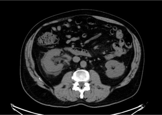
DOI: https://doi.org/10.21980/J8GD2TThe CT scan demonstrates nephrolithiasis with associated forniceal rupture. Encircled in the yellow outline is fluid, demonstrating a forniceal rupture. The stone is in the proximal aspect of the ureter, as highlighted by the purple arrow.


