Open Globe with Intraocular Foreign Body
History of present illness:
A 7-year-old male with no past medical history presented to the emergency department with 24 hours of right eye pain and redness. The previous day he had been smashing rocks on the floor and felt an object strike his eye. The following morning, he was seen by his pediatrician who referred him to an ophthalmologist. No same-day appointments were available, so the family sought emergency care. On arrival, he described right eye pain which increased with light exposure and blurry vision.
Significant findings:
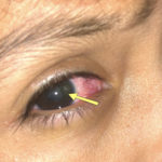
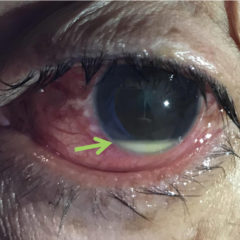
On physical exam, his extraocular movements were intact. The right anterior chamber appeared cloudy, particularly nasal to the pupil. The conjunctiva of the right eye was injected. The right pupil was 3 mm and sluggishly reactive and appeared slightly irregular (see yellow arrow). Of note, the right eye also had a 1 mm hypopyon, indicating inflammation of the anterior chamber, which was visible on slit lamp examination (not pictured). There was no fluorescein uptake or Seidel sign. His visual acuity was 20/60 OD (right eye) and 20/20 OS (left eye).
Computed tomography (CT) of the orbits was performed without contrast. There was a spherical high-density foreign body in the right posterior globe, inferiorly, at the 5 o’clock position measuring 3mm in diameter. No hematoma, lens dislocation, or significant decrease in volume was noted.
Discussion:
An open globe due to a small intraocular foreign body (IOFB) can be a challenging diagnosis. Findings that aided in diagnosis in this case included a slightly irregular pupil, clouding of the anterior chamber and a small hypopyon which may have indicated early onset of endophthalmitis. Careful history and physical examination, combined with CT are the ideal diagnostic modalities for patients presenting with an open globe secondary to an IOFB.1
The physical exam typically includes fluorescein examination with a Wood’s lamp. The Seidel Test is performed by applying fluorescein and observing for dye streaming away from the puncture site by leaking aqueous humor, indicative of penetrating trauma. However, it is important to recognize that the Seidel sign has sensitivity of just 70%.2 Ultrasound, used with caution (large amounts of gel, minimal pressure), may be used to evaluate for IOFB in compliant patients, and may also identify other pathologies such as retinal detachment and vitreous hemorrhage.3-5 If an open globe injury is diagnosed or highly suspected, tonometry should be avoided to prevent further extruding ocular contents.5
Open globe injuries should receive systemic intravenous antibiotics to reduce the chance of developing endophthalmitis.6 Retained intraocular foreign bodies are associated with endophthalmitis in 7% to 12% of cases.7 The association is reduced to 0.9% with 48 hours of intravenous antibiotics and prompt ophthalmology intervention.8
In this case, intravenous cefepime and vancomycin were given in the emergency department. Ophthalmology was consulted and their exam noted a 3 mm laceration nasal to the pupil, intermittent Seidel sign and early cataract formation in the right eye. The patient then underwent emergent operative repair.
Topics:
Open globe, intraocular foreign body, CT of the orbits, endophthalmitis.
References:
- Parke DW, Flynn HW, Fisher YL. Management of intraocular foreign bodies: a clinical flight plan. Can J Ophthalmol. 2013;48(1):8-12. doi: 10.1016/j.jcjo.2012.11.005
- Hoffstetter P, Schreyer AG, Schreyer CI, et al. Multidetector CT (MD-CT) in the diagnosis of uncertain open globe injuries. Rofo. 2010;182(2):151-154. doi: 10.1055/s-0028-1109659
- Andreoli MT, Yiu G, Hart L, Andreoli CM. B-scan ultrasonography following open globe repair. Eye (Lond). 2014;28(4):381-385. doi: 10.1038/eye.2013.289
- Lecler A, Pinel A, Koskas P. Open globe injury: ultrasound first! Am J Neuroradiol. 2017;38(11):E99-E100. doi: 10.3174/ajnr.A5282
- Li X, Zarbin MA, Bhagat N. Pediatric open globe injury: a review of the literature. J Emerg Trauma Shock. 2015;8(4):216-223. doi: 10.4103/0974-2700.166663
- Ahmed Y, Schimel AM, Pathengay A, Colyer MH, Flynn HW. Endophthalmitis following open-globe injuries. Eye (Lond). 2012;26(2):212-217. doi: 10.1038/eye.2011.313
- Mieler WF, Ellis MK, Williams DF, Han DP. Retained intraocular foreign bodies and endophthalmitis. Ophthalmology. 1990;97(11):1532-1538. doi: 10.1016/S0161-6420(90)32381-3
- Andreoli CM, Andreoli MT, Kloek CE, Ahuero AE, Vavvas D, Durand ML. Low rate of endophthalmitis in a large series of open globe injuries. Am J Ophthalmol. 2009;147(4):601-608.e2. doi: 10.1016/j.ajo.2008.10.023


