Ultrasound
Evaluation of Snake Bites with Bedside Ultrasonography
DOI: https://doi.org/10.21980/J84S7DHistory of present illness: While watering his lawn, a 36-year-old man felt two sharp bites to his bilateral ankles. He reports that he then saw a light brown, 2-foot snake slither away from him. He came to the emergency department because of pain and swelling in his ankles and inability to bear weight. Physical examination revealed bilateral ankle swelling and
Glass Foreign Body Hand Radiograph
DOI: https://doi.org/10.21980/J8W92HHistory of present illness: A 27-year-old female sustained an injury to her left hand after she tripped and fell on a vase. She presented to the emergency department (ED) complaining of pain over the laceration. Upon examination, patient presented with multiple small abrasions of the medial aspect of the left 5thdigit that are minimally tender. Additionally, she has one 0.5cm
Point-of-care Ultrasound for the Diagnosis of a Gluteal Abscess
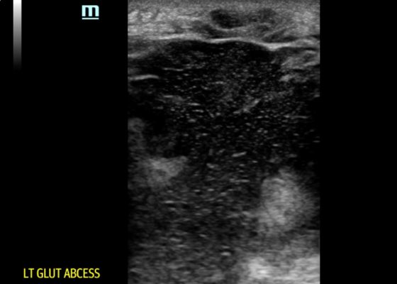
DOI: https://doi.org/10.21980/J8VH1WPOCUS reveals a large, hypoechoic soft tissue abscess with debris and tracks extending to the bottom of the image. Furthermore, when compressed, movement of the abscess contents is appreciated. There is also superficial cobble-stoning consistent with overlying cellulitis and soft tissue edema.
Point-of-care Ultrasound Detection of Endophthalmitis
DOI: https://doi.org/10.21980/J8G634The patient’s ultrasound revealed an attached retina and a complex network of hyperechoic, mobile, membranous material in the posterior segment.
Bedside Ultrasound for the Diagnosis of Peritonsillar Abscess
DOI: https://doi.org/10.21980/J8N33NThe first video is an intraoral ultrasound using the high frequency endocavitary probe demonstrating an anechoic fluid collection adjacent to the patient’s enlarged left tonsil. The second video shows real-time ultrasound-guided successful drainage of the PTA.
Point-of-care Ultrasound for the Diagnosis of Ectopic Pregnancy
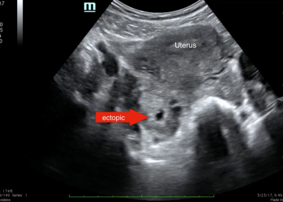
DOI: https://doi.org/10.21980/J8VK7VThe transabdominal pelvic ultrasound shows an empty uterus (annotated) with free fluid and a right sided extrauterine gestational sac representing an ectopic pregnancy (red arrow).
Introducing point-of-care ultrasound through competency-based simulation education using a fractured chicken bone model
DOI: https://doi.org/10.21980/J8GG95To introduce medical students to PoCUS with an inexpensive, reproducible, and educationally effective model using fractured chicken bones set in gelatin, and to assess medical students’ abilities to identify simulated long-bone fractures using PoCUS.
Using Bedside Ultrasound to Rapidly Differentiate Shock
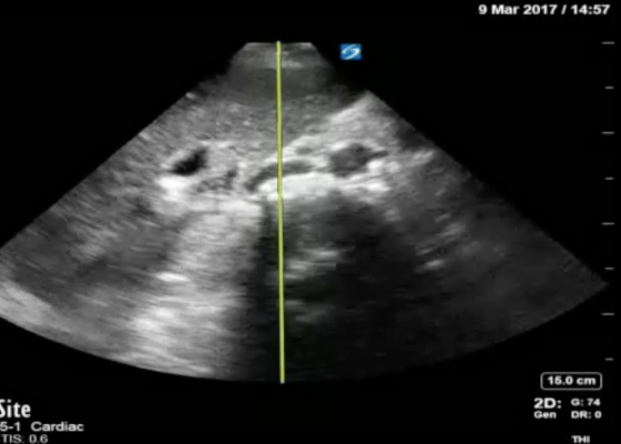
DOI: https://doi.org/10.21980/J8S047A RUSH exam demonstrated hyperdynamic cardiac contractility and collapse of the inferior vena cava (IVC) with probe compression more than 50% suggesting hypovolemia likely secondary to sepsis. Incidentally, Morrison’s pouch revealed a large right renal cyst but no signs of free fluid. A computed tomography of abdomen/pelvis showed a 10.8 x 9.5 cm right renal cyst and left lower lobe pneumonia.


