• Registration
  • Login
JETem
  • Home
  • About
    • Aim and Scope
    • Our Team
    • Editorial Board
    • FAQ
  • Issues
    • Current Issue
    • Ahead of Print
    • Past Issues
  • Visual EM
    • Latest Visual EM
    • Search Visual EM
    • Thumbnail Library
  • For Authors
    • Instructions for Authors
    • Submit to JETem
    • Photo Consent
    • Policies
      • Peer Review Policy
      • Copyright Policy
      • Editorial Policy, Ethics and Responsibilities
      • Conflicts of Interest & Informed Consent
      • Open Access Policy
  • For Reviewers
    • Instructions for JETem Reviewers
    • Interested in Being a JETem Reviewer?
  • Topic
    • Abdominal / Gastroenterology
    • Administration
    • Board Review
    • Cardiology / Vascular
    • Clinical Informatics, Telehealth and Technology
    • Dermatology
    • EMS
    • Endocrine
    • ENT
    • Faculty Development
    • Genitourinary
    • Geriatrics
    • Hematology / Oncology
    • Infectious Disease
    • Miscellaneous
    • Neurology
    • Ob / Gyn
    • Ophthalmology
    • Orthopedics
    • Pediatrics
    • Procedures
    • Psychiatry
    • Renal / Electrolytes
    • Respiratory
    • Toxicology
    • Trauma
    • Ultrasound
    • Wellness
    • Wilderness
  • Modality
    • Curricula
    • Innovations
    • Lectures
    • Oral Boards
    • Podcasts
    • Simulation
    • Small Group Learning
    • Team Based Learning
    • Visual EM
  • Contact Us

Cholelithiasis: WES Sign

Hamid Ehsani-Nia, MS* and Shannon Toohey, MD, MAEd^

*Rowan University School of Osteopathic Medicine, Stratford, NJ
^University of California, Irvine, Department of Emergency Medicine, Orange, CA

Correspondence should be addressed Hamid Ehsani-Nai at hamidnia101@gmail.com

DOI: https://doi.org/10.21980/J8X300Issue 2:1
Abdominal/GastroenterologyUltrasoundVisual EM
No ratings yet.

History of present illness:

A 33-year-old female with multiple previous episodes of biliary colic presented to the emergency department with severe right upper quadrant pain that started 30 minutes prior to arrival. She characterized her pain as sharp and pulling and radiating to the back. Upon physical examination, patient was in acute distress with severe tenderness to right upper quadrant palpation. Her abdomen was soft, with no guarding or rebound tenderness.

Significant findings:

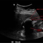
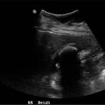
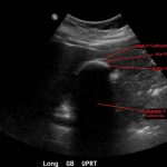
Abdominal ultrasound showed the classic presentation of the Wall-Echo-Shadow (WES) sign. The superficial aspect of the gallbladder wall is represented by a hyperechogenic curve. Below this, bile fluid is represented by hypoechogenicity. Underneath the bile fluid is the echo of the dense border created by the collection of gallstones, represented by a hyperechogenic curve. Due to the high density of the gallstones, nothing deeper can be visualized (including other gallstones or the far end of the gallbladder); this is the shadow.

Discussion:

The WES sign, first described in the early 1980s, is a clear way of identifying cholelithiasis when the condition has progressed to fill the entire lumen of the gallbladder.1 In this case, the lumen of the gallbladder looks hypoechogenic relative to the surrounding tissue, much like an empty gallbladder. This is due to the border of the dense stones opposing the near wall of the gallbladder casting a shadow over everything distal to the stones. These key visualization techniques decrease non-visualization of chronic cholelithiasis.2,3,4 In most cases, WES sign is sufficient to diagnose cholelithiasis. Of note, “porcelain gallbladder” or collapsed duodenum can have the potential produce similar ultrasonographic findings.4,5

Topics:

Ultrasound, gallbladder, WES, wall echo shadow, cholelithiasis, gallstones, abdominal pain.

References:

  1. Macdonald FR, Cooperberg PL, Cohen MM. The WES triad – a specific sonographic sign of gallstones in the contracted gallbladder. Gastrointest Radiol. 1981;6(1):39-41. doi: 10.1007/BF01890219
  2. George N, Dawkins A, Disantis D. The wall-echo-shadow (WES) sign. Abdom Imaging. 2015;40(7):2903. doi: 10.1007/s00261-015-0401-4
  3. Raptopoulos V, D’orsi C, Smith E, Reuter K, Moss L, Kleinman P. Dynamic cholecystosonography of the contracted gallbladder: the double-arc-shadow sign. AJR Am J Roentgenol. 1982;138(2):275-278. doi: 10.2214/ajr.138.2.275
  4. Rybicki FJ. The WES sign. Radiology. 2000;214(3):881-882. doi: 10.1148/radiology.214.3.r00mr38881
  5. Solomon DM, Paippattuthara J, Zoberman H, Hahn B. Images in emergency medicine: wall-echo-shadow (WES) complex. J Emerg Med. 2012;43(3):e203-204. doi: 10.1016/j.jemermed.2011.01.001
Icon

WES Sign - Case Report

1 file(s) 376 KB
Download
Icon

WES Sign - Images

1 file(s) 1.46 MB
Download
Issue 2:1Ultrasound

Reviews:

No ratings yet.

Please rate this


A Formalized Three-Year Emergency Medicine...

23 Jan, 17

Monteggia Fracture in an Assault Patient

23 Jan, 17
JETem is an online, open access, peer-reviewed journal-repository for EM educators

Most Viewed

  • Telemedicine Consult for Shortness of Breath Due to Sympathetic Crashing Acute Pulmonary Edema
  • Anticholinergic Toxicity in the Emergency Department
  • The Suicidal Patient in the Emergency Department Team-Based Learning Activity
  • Child Maltreatment Education: Utilizing an Escape Room Activity to Engage Learners on a Sensitive Topic
  • Acute Chest Syndrome

Visit Our Collaborators

About

Education

Learners should benefit from active learning. JETem accepts submissions of team-based learning, small group learning, simulation, podcasts, lectures, innovations, curricula, question sets, and visualEM.

Scholarship

We believe educators should advance through the scholarship of their educational work. JETem gives educators the opportunity to publish scholarly academic work so that it may be widely distributed, thereby increasing the significance of their results.

Links

  • Home
  • Aim and Scope
  • Current Issue
  • For Reviewers
  • Instructions for Authors
  • Contact Us

Newsletter

Sign up to receive updates from JETem regarding newly published issues and findings.

Copyright © 2016 JETem. All rights reserved.