• Registration
  • Login
JETem
  • Home
  • About
    • Aim and Scope
    • Our Team
    • Editorial Board
    • FAQ
  • Issues
    • Current Issue
    • Ahead of Print
    • Past Issues
  • Visual EM
    • Latest Visual EM
    • Search Visual EM
    • Thumbnail Library
  • For Authors
    • Instructions for Authors
    • Submit to JETem
    • Photo Consent
    • Policies
      • Peer Review Policy
      • Copyright Policy
      • Editorial Policy, Ethics and Responsibilities
      • Conflicts of Interest & Informed Consent
      • Open Access Policy
  • For Reviewers
    • Instructions for JETem Reviewers
    • Interested in Being a JETem Reviewer?
  • Topic
    • Abdominal / Gastroenterology
    • Administration
    • Board Review
    • Cardiology / Vascular
    • Clinical Informatics, Telehealth and Technology
    • Dermatology
    • EMS
    • Endocrine
    • ENT
    • Faculty Development
    • Genitourinary
    • Geriatrics
    • Hematology / Oncology
    • Infectious Disease
    • Miscellaneous
    • Neurology
    • Ob / Gyn
    • Ophthalmology
    • Orthopedics
    • Pediatrics
    • Pharmacology
    • Procedures
    • Psychiatry
    • Renal / Electrolytes
    • Respiratory
    • Social Determinants of Health
    • Toxicology
    • Trauma
    • Ultrasound
    • Urology
    • Wellness
    • Wilderness
  • Modality
    • Curricula
    • Innovations
    • Lectures
    • Oral Boards
      • Structured Interview
      • Communication Case
    • Podcasts
    • Simulation
    • Small Group Learning
    • Team Based Learning
    • Visual EM
  • Contact Us

Neurology

Creative Commons images

Viridans streptococci Intracranial Abscess Masquerading as Metastatic Disease

Brandon Ruderman, MD*, Traci Thoureen, MD* and Joshua Broder, MD*

DOI: https://doi.org/10.21980/J8CH05 Issue 3:1 No ratings yet.
A non-contrast CT (Figure 1) revealed a large hypoattenuating left parietal lesion. When the CT was enhanced with intravenous contrast (Figure 2), the same lesion showed peripheral rim enhancement, suggestive of a brain abscess.
Infectious DiseaseNeurologyVisual EM
Creative Commons images

Dense MCA Sign

Richard J Chen, MD* and Grant Wei, MD*

DOI: https://doi.org/10.21980/J8CS66Issue 2:3 No ratings yet.
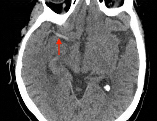
A non-contrast computed tomography (CT) scan showed a hyperdensity along the right middle cerebral artery (MCA) consistent with acute thrombus. The red arrow highlights the hyperdensity in the annotated image.
NeurologyVisual EM

Status Epilepticus in the Emergency Department

Jonathan Lee, BS* and Alisa Wray, MD*

DOI: https://doi.org/10.21980/J8RC7VIssue 2:2 No ratings yet.
At the end of this simulation session, the learner will: 1) Demonstrate the management of status epilepticus 2) Justify when airway intervention is needed for status epilepticus 3) Describe risk factors for status epilepticus 4) Prepare a differential diagnosis for the causes in status epilepticus.
NeurologySimulation
Creative Commons images

Presentation of Significant Subarachnoid Hemorrhage without Loss of Consciousness

Nicholas Taylor, BA* and Shannon Toohey, MD, MAEd*

DOI: https://doi.org/10.21980/J80W29 Issue 2:2 No ratings yet.
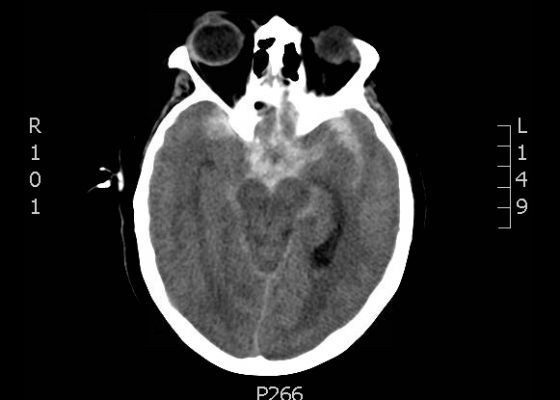
A non-contrast head CT demonstrated extensive subarachnoid hemorrhage occupying both cerebral convexities, the anterior interhemispheric fissure, the sylvian fissures, and the basal cisterns. Later CTA would show an 8 mm by 7 mm by 8mm MCA aneurysm near the M1/M2 junction and two pericallosal artery aneurysms, 7 by 6 mm and 8 by 5 mm respectively.
NeurologyVisual EM
Creative Commons images

Acute Subdural Hematoma

Ellen Lester, BS*, Jonathan Peña, MD* and Warren Wiechmann, MD, MBA*

DOI: https://doi.org/10.21980/J87C76 Issue 2:2 No ratings yet.
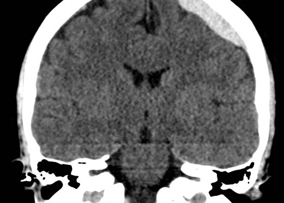
Non-contrast Computed Tomography (CT) of the Head showed a dense extra-axial collection along the left frontal and parietal regions, extending superior to the vertex with mild mass effect, but no midline shift.
NeurologyVisual EM
Creative Commons images

Intracranial Hemorrhage Following a 3-week Headache

John Jiao, MHS* and Alisa Wray, MD*

DOI: https://doi.org/10.21980/J89885 Issue 2:1 No ratings yet.
The patient’s head CT showed a significant area of hyperdensity consistent with an intracranial hemorrhage located within the left frontal parietal lobe (red arrow). Additionally, there is rightward midline shift up to 1.1cm (green arrow) and entrapment of the right lateral ventricle (blue arrow).
NeurologyVisual EM

Approach to Acute Headache: A Flipped Classroom Module for Emergency Medicine Trainees

Jeff Riddell, MD*, Stacy Sawtelle, MD^, Paul Jhun, MD‡, James Comes, MD^, Ramin Tabatabai, MD~, Daniel Joseph, MD~, Jan Shoenberger, MD~, Esther Chen, MD‡, Christopher Fee, MD‡, and Stuart Swadron, MD~

DOI: https://doi.org/10.21980/J8WC73Issue 1:1 No ratings yet.
At the end of this module, the learner will be able to: 1) list the diagnoses critical to the emergency physician that may present with headache; 2) identify key historical and examination findings that help differentiate primary (benign) from secondary (serious) causes of headache; 3) discuss the indications for diagnostic imaging, lumbar puncture and laboratory testing in patients with headache; 4) recognize life-threatening diagnoses on CT imaging and CSF examination; 5) describe treatment strategies to relieve headache symptoms.
NeurologySmall Group Learning
Creative Commons images

Normal Head CT

No ratings yet.
  Keywords: radiology, normal, CT head, neuro, axial
Neurology
«‹4
Page 4 of 4

JETem is an online, open access, peer-reviewed, journal-repository for EM educators. We are PMC Indexed.

Most Viewed

  • The Silent Saboteur: Teaching the Clinical Implications of Occult Hypoxemia & Social Determinants of Health via a Pulmonary Embolism Case
  • Diabetic Ketoacidosis and Necrotizing Soft Tissue Infection
  • My Broken Heart
  • Stabilization of Cardiogenic Shock for Critical Care Transport, a Simulation
  • Innovative Ultrasound-Guided Erector Spinae Plane Nerve Block Model for Training Emergency Medicine Physicians

Visit Our Collaborators

Creative Commons Licence
This work is licensed under a Creative Commons Attribution 4.0 International License.

About

Education

Learners should benefit from active learning. JETem accepts submissions of team-based learning, small group learning, simulation, podcasts, lectures, innovations, curricula, question sets, and visualEM.

Scholarship

We believe educators should advance through the scholarship of their educational work. JETem gives educators the opportunity to publish scholarly academic work so that it may be widely distributed, thereby increasing the significance of their results.

Links

  • Home
  • Aim and Scope
  • Current Issue
  • For Reviewers
  • Instructions for Authors
  • Contact Us

Newsletter

Sign up to receive updates from JETem regarding newly published issues and findings.

Copyright Creative Commons Attribution 4.0 International