Dermatology
Open Chest Wound with Sternal Fracture in the Emergency Department, a Case Report
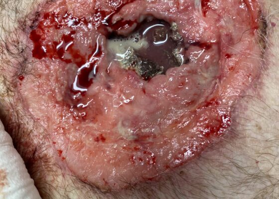
DOI: https://doi.org/10.5070/M5.52202The image demonstrates the large chronic-appearing wound of the patient’s anterior chest as well as the visible fractured segments of the patient’s exposed sternum. The sternum is necrotic appearing concerning for a chronic process including osteomyelitis and malignancy. Purulent drainage is visible on the wound consistent with an infectious process.
Effects of Volume Overload: A Case Report of an Edema Bulla
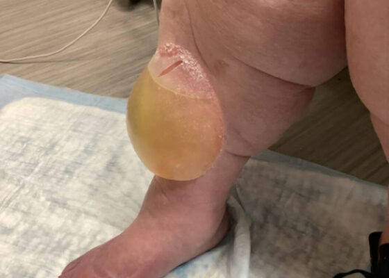
DOI: https://doi.org/10.5070/M5.52206This image shows a large edema bulla on the patient's right shin. The bulla is 10 x 10 cm, filled with serous fluid, has a spontaneously occurring defect in the skin of the superior portion of the bulla, and is non-erythematous. The bulla is much larger than the 1-5 cm edema bullae described in the literature. As edema bulla is primarily a clinical diagnosis, taking the full history and physical exam into account is essential to recognize these bullae.
Case Report of a Dermatologic Reaction to Wound Closure Strips and Liquid Adhesive
DOI: https://doi.org/10.21980/J8.52256The patient removed the splint, and the wound were notable for erythematous bullae (blue arrow), blisters (yellow arrow), and skin maceration (red arrow) in the distribution under the wound closure strips. Of note, there was no surrounding erythema with poorly defined borders.
Metastatic Calcinosis Cutis in the Emergency Department: A Case Report
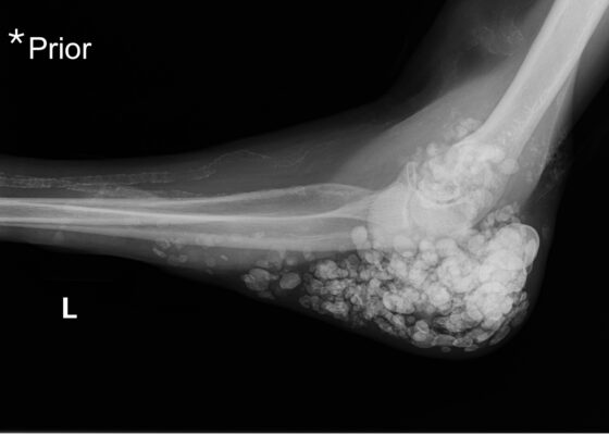
DOI: https://doi.org/10.21980/J87Q00X-ray imaging was obtained of the left elbow and showed soft tissue calcium deposits. Radiology stated, “massive periarticular calcinosis of renal failure obscures fine osseous detail. Several of the largest calcifications have decompressed since the prior exam and may contribute to the drainage observed clinically. Superimposed infection is not excluded.” X-rays with an asterisk are the comparison images from two months previous to the visit. Areas of decompression are highlighted in blue demonstrating that some of the larger calcified nodules are no longer present.
A Case Report of Calciphylaxis
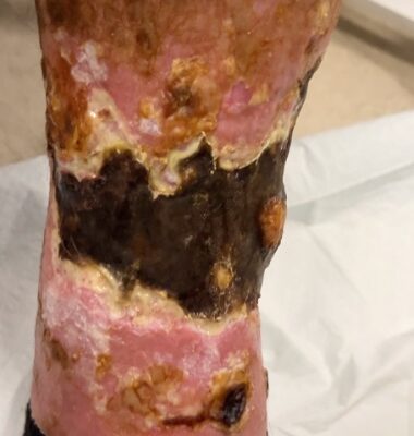
DOI: https://doi.org/10.21980/J8KW8VOn arrival for this visit, the patient was nontoxic appearing with stable vital signs. The physical exam was notable for deep, ulcerated, bilateral anterior leg wounds with purulent drainage and large areas of eschar (see photographs).
A Case Report on an Elusive Incident of Erythema Multiforme
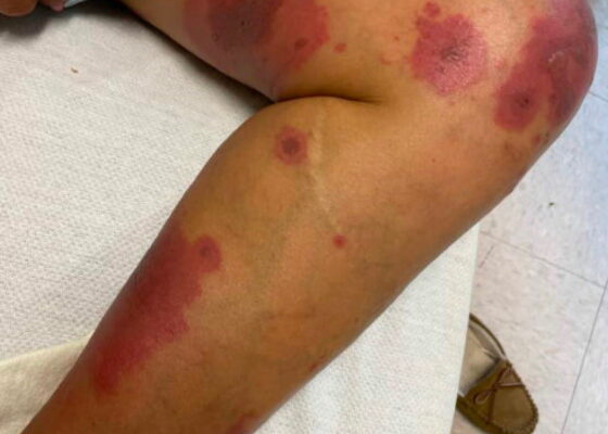
DOI: https://doi.org/10.21980/J8BM0WHer physical exam was notable for multiple scattered tense vesicles on an erythematous base along the left and right lower extremities and right upper extremity. The lesions were excoriated and in different stages of evolution. No oral, mucosal, or conjunctival lesions were found. Physical exam was otherwise unremarkable.
A Case Report on Dermatomyositis in a Female Patient with Facial Rash and Swelling
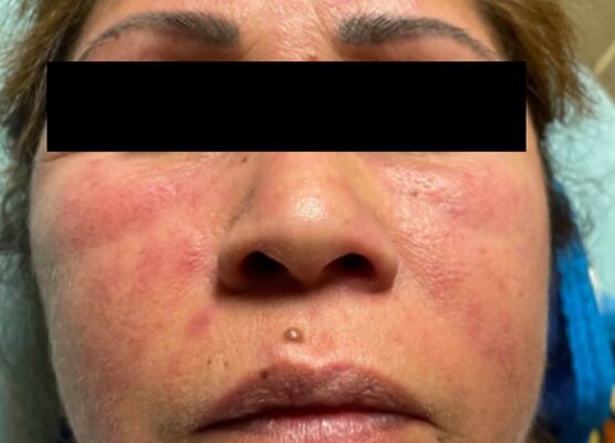
DOI: https://doi.org/10.21980/J8506DThe physical exam revealed significant periorbital swelling, facial edema, and a maculopapular rash across the upper chest, symmetrically across the extensor surfaces of the hands and the bilateral arms and thighs. The photograph of her face shows light-red to violaceous macules and patches, with inclusion of the nasolabial folds as well the forehead and upper eyelids with periorbital edema (heliotrope sign). The other rash images show “Shawl sign” (photograph of back showing erythema over the posterior aspect of the upper back), V sign (photograph of chest showing light-red violaceous plaque on mid-chest), Gottron's papules (photograph of hands showing light red scaly papules overlying the right proximal interphalangeal joint [R PIP] and the metacarpophalangeal joint [MCP], and holster sign (photograph of thigh showing light red patches on bilateral lateral thighs). This distribution of rashes is pathognomonic for DM.
A Case Report of Dermatographia
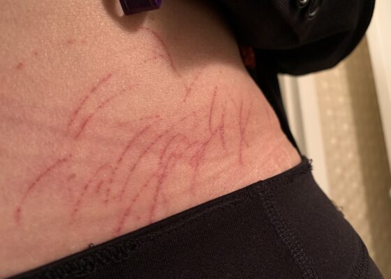
DOI: https://doi.org/10.21980/J8P05PPhysical examination was unremarkable except for the urticaria on the right aside of her abdomen (white arrow) with overlying excoriations (stars). Of note, there were no burrows, papules or vesicles in the typical locations including the webs of the fingers, wrists, axillae, areolae, or genitalia. Examination of the linear dermatographia clearly revealed superficial wheals, versus underlying serpiginous lesions.

