• Registration
  • Login
JETem
  • Home
  • About
    • Aim and Scope
    • Our Team
    • Editorial Board
    • FAQ
  • Issues
    • Current Issue
    • Ahead of Print
    • Past Issues
  • Visual EM
    • Latest Visual EM
    • Search Visual EM
    • Thumbnail Library
  • For Authors
    • Instructions for Authors
    • Submit to JETem
    • Photo Consent
    • Policies
      • Peer Review Policy
      • Copyright Policy
      • Editorial Policy, Ethics and Responsibilities
      • Conflicts of Interest & Informed Consent
      • Open Access Policy
  • For Reviewers
    • Instructions for JETem Reviewers
    • Interested in Being a JETem Reviewer?
  • Topic
    • Abdominal / Gastroenterology
    • Administration
    • Board Review
    • Cardiology / Vascular
    • Clinical Informatics, Telehealth and Technology
    • Dermatology
    • EMS
    • Endocrine
    • ENT
    • Faculty Development
    • Genitourinary
    • Geriatrics
    • Hematology / Oncology
    • Infectious Disease
    • Miscellaneous
    • Neurology
    • Ob / Gyn
    • Ophthalmology
    • Orthopedics
    • Pediatrics
    • Pharmacology
    • Procedures
    • Psychiatry
    • Renal / Electrolytes
    • Respiratory
    • Social Determinants of Health
    • Toxicology
    • Trauma
    • Ultrasound
    • Urology
    • Wellness
    • Wilderness
  • Modality
    • Curricula
    • Innovations
    • Lectures
    • Oral Boards
      • Structured Interview
      • Communication Case
    • Podcasts
    • Simulation
    • Small Group Learning
    • Team Based Learning
    • Visual EM
  • Contact Us

Abdominal/Gastroenterology

Creative Commons images

Bedside Ultrasound for the Diagnosis of Small Bowel Obstruction

Alexander Anshus, BS* and Maili Alvarado, MD^

DOI: https://doi.org/10.21980/J86W6PIssue 2:4 No ratings yet.
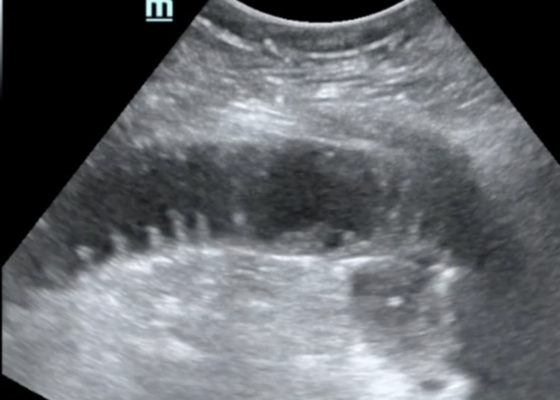
The POCUS utilizing the low frequency curvilinear probe demonstrates fluid-filled, dilated bowel loops greater than 2.5cm with to-and-fro peristalsis, and thickened bowel walls greater than 3mm, concerning for SBO. 
Abdominal/GastroenterologyVisual EM
Creative Commons images

Choledocholithiasis

Jonathan Peña, MD*

DOI: https://doi.org/10.21980/J8Q62X Issue 2:3 No ratings yet.
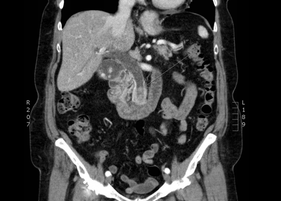
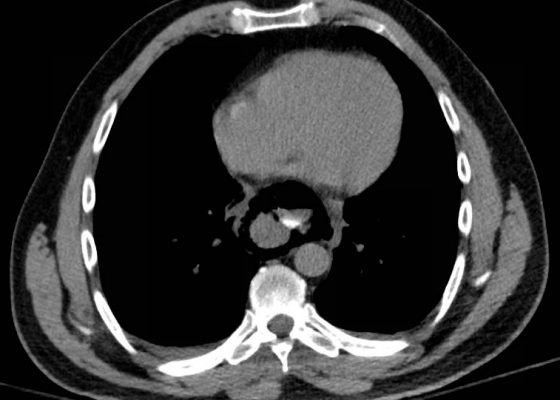
Computed tomography (CT) was significant for two large gallstones measuring 1.1 centimeters impacted at the level of the pancreatic head with associated common bile duct (CBD) dilatation.
Abdominal/GastroenterologyVisual EM
Creative Commons images

Volvulus

Sari Lahham, MD, MBA*, Kathryn Bennett, BS* and Mohammad Helmy, MD†

DOI: https://doi.org/10.21980/J8JH0Q Issue 2:3 No ratings yet.
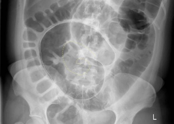
Upright and supine frontal radiographs of the abdomen demonstrate gas dilation of the large bowel from the level of the cecum to the sigmoid colon with air fluid levels (yellow arrows). There is a swirled configuration of the distal descending to sigmoid colon indicating the level of the volvulus (dashed yellow line) and giving rise to the classic “coffee bean” sign (dotted white tracing). Note the elevated left hemidiaphragm on the upright view reflecting abdominal distention with increased intra-abdominal pressure (red arrow).
Abdominal/GastroenterologyVisual EM
Creative Commons images

Esophageal Perforation

Valentina Park, BS* and Jason Mefford, MD^

DOI: https://doi.org/10.21980/J8K91B Issue 2:3 No ratings yet.
History of present illness: A 51-year-old male with history of gastroesophageal reflux disease status post multiple endoscopies presented to the emergency department with severe abdominal pain. Paramedics reported the patient appeared diaphoretic on arrival and maintained stable vital signs during transit. The patient reported taking Prilosec that morning before eating breakfast, after which he felt like something was stuck in
Abdominal/GastroenterologyVisual EM
Creative Commons images

Perforated Gastric Ulcer with Intra-abdominal Abscess

Leslie Palmerlee, MD, MPH*, Scott Mackey, DO* and Michael Petrauskis, MD, MEd*

DOI: https://doi.org/10.21980/J82H0C Issue 2:3 No ratings yet.
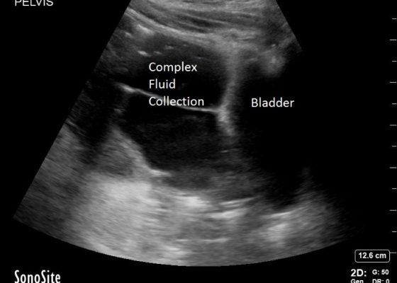
Bedside ultrasound revealed a large volume of free fluid in the right upper quadrant and in the pelvis. The fluid appeared complex with multiple septations. Its appearance was not consistent with ascites or acute intra-abdominal free fluid due to striations and pockets.
Abdominal/GastroenterologyVisual EM
Creative Commons images

Bowel Perforation complicating an incarcerated inguinal hernia

Adam Sigal, MD* and Jamie Slotkin, DO^

DOI: https://doi.org/10.21980/J8D30BIssue 2:2 No ratings yet.
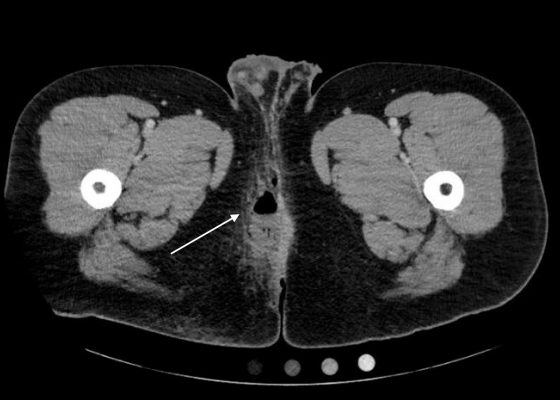
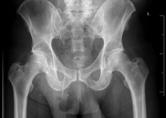
The AP and lateral pelvis x-rays revealed two sewing needles, 60 mm in length, within the soft tissue over the anterior right lower hemipelvis. In addition, the AP view showed emphysema involving the right hemiscrotum (arrow), concerning for perforated bowel.
Abdominal/GastroenterologyVisual EM
Creative Commons images

A Toddler with Abdominal Pain and Emesis

Saema Said, BS* and Kevin Koenig, MD*

DOI: https://doi.org/10.21980/J8XW2P Issue 2:2 No ratings yet.
In the long axis video, the appendix appears as an enlarged, non-compressible, blind-ending tubular structure (white arrow) with distinct appendiceal wall layers and lack of peristalsis. In the short axis video, the appendix appears as a target sign (yellow arrow) between the abdominal and psoas muscles. The maximal outer diameter (MOD) measures 11.8mm and the appendix wall measures 0.17mm. There is trace adjacent free fluid and echogenic periappendiceal fat. Transverse axis video and image (red arrow) demonstrate that the appendix is not compressible. These findings are consistent with acute appendicitis.
Abdominal/GastroenterologyInfectious DiseasePediatricsUltrasoundVisual EM

Novel Emergency Medicine Curriculum Utilizing Self-Directed Learning and the Flipped Classroom Method: Gastrointestinal Emergencies Small Group Module

Andrew King, MD, FACEP*, Elizabeth Matheson^, BS, Christopher San Miguel, MD*, Sarah Greenberger, MD*, Michael Barrie, MD*, Jillian McGrath, MD*, Howard Werman, MD*, Ashish Panchal, MD*, Daniel Martin, MD*, David P Bahner, MD*, Sorabh Khandelwal, MD* and Jennifer Mitzman, MD*

DOI: https://doi.org/10.21980/J8MS37 Issue 2:1 No ratings yet.
We aim to teach the presentation and management of GI emergencies through the creation of a flipped classroom design. This unique, innovative curriculum utilizes resources chosen by education faculty and resident learners, study questions, real-life experiences, and small group discussions in place of traditional lectures. In doing so, a goal of the curriculum is to encourage self-directed learning, improve understanding and knowledge retention, and improve the educational experience of our residents.
Abdominal/GastroenterologyCurriculaSmall Group Learning
Creative Commons images

Ventriculoperitoneal Shunt Migration

Justin P Puller, MD* and Jonathan T Miller, MD*

DOI: https://doi.org/10.21980/J8G019Issue 2:1 No ratings yet.
An immediate post-op abdominal x-ray performed after the patient’s VP shunt revision 30 days prior to this ED visit reveals the VP shunt tip in the mid abdomen. A CT of the abdomen performed on the day of the ED visit reveals the VP shunt tip interposed between the spleen and the diaphragm.
Abdominal/GastroenterologyVisual EM
Creative Commons images

Computed Tomography Diagnosis of Appendicitis

Christopher Libby, MPH* and Shannon Toohey, MD, MAEd^

DOI: https://doi.org/10.21980/J8F30NIssue 2:1 No ratings yet.
The CT abdomen/pelvis with IV contrast shows a dilated appendix (see red outline) with thickened, hyperenhancing wall (see blue outline) best visualized in the axial and coronal planes.
Abdominal/GastroenterologyVisual EM
«‹6›
Page 6 of 7

JETem is an online, open access, peer-reviewed, journal-repository for EM educators. We are PMC Indexed.

Most Viewed

  • The Silent Saboteur: Teaching the Clinical Implications of Occult Hypoxemia & Social Determinants of Health via a Pulmonary Embolism Case
  • Diabetic Ketoacidosis and Necrotizing Soft Tissue Infection
  • My Broken Heart
  • Stabilization of Cardiogenic Shock for Critical Care Transport, a Simulation
  • Innovative Ultrasound-Guided Erector Spinae Plane Nerve Block Model for Training Emergency Medicine Physicians

Visit Our Collaborators

Creative Commons Licence
This work is licensed under a Creative Commons Attribution 4.0 International License.

About

Education

Learners should benefit from active learning. JETem accepts submissions of team-based learning, small group learning, simulation, podcasts, lectures, innovations, curricula, question sets, and visualEM.

Scholarship

We believe educators should advance through the scholarship of their educational work. JETem gives educators the opportunity to publish scholarly academic work so that it may be widely distributed, thereby increasing the significance of their results.

Links

  • Home
  • Aim and Scope
  • Current Issue
  • For Reviewers
  • Instructions for Authors
  • Contact Us

Newsletter

Sign up to receive updates from JETem regarding newly published issues and findings.

Copyright Creative Commons Attribution 4.0 International