Posts by JETem
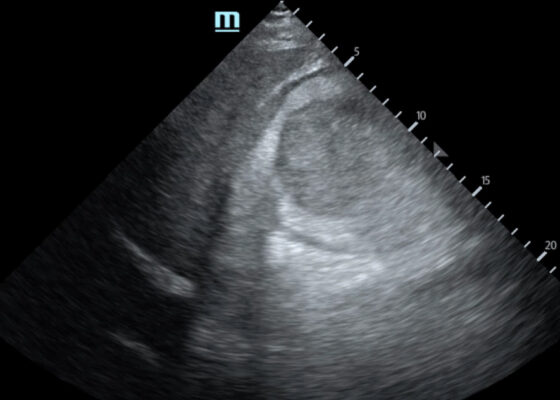
Trauma by Couch: A Case Report of a Massive Traumatic Retroperitoneal Hematoma
DOI: https://doi.org/10.21980/J84D2QUpon arrival at the trauma center, a FAST revealed a large, well-circumscribed abnormality (red outline) deep to the liver (blue outline and star) and gallbladder (green outline and star). The right kidney and hepatorenal space were not clearly visualized. The remainder of the FAST showed no free fluid in the splenorenal space, pelvis, and no pericardial effusion. He had lung sliding bilaterally.
A Case Report of Invasive Mucormycosis in a COVID-19 Positive and Newly-Diagnosed Diabetic Patient
DOI: https://doi.org/10.21980/J81M1GOn physical exam, when the patient was asked to try and look to her right, the right eye failed to move laterally/abduct (blue arrow). Additionally, when asked to look straight ahead, the eye was slightly adducted (red arrow). There was a lack of motion of the right eye in abduction when the patient was asked to look to her right (yellow arrow).
A Patient with Generalized Weakness – A Case Report
DOI: https://doi.org/10.21980/J8593CThe CT of the abdomen and pelvis showed evidence of a large subcapsular rim-enhancing fluid collection with multiple gas and air-fluid levels along the right kidney measuring 8 x 4 cm axially and 11 cm craniocaudally (blue outline) with mass effect on the right renal parenchyma (yellow outline). Another suspected fluid collection adjacent to the upper pole of the right kidney measuring 4 x 3.4 cm was noted (red outline). Bilateral pyelonephritis was suggested without hydronephrosis or nephrolithiasis. The findings suggested complicated pyelonephritis with emphysematous abscess and hematoma formation.
How to Build a Low-Cost Video-Assisted Laryngoscopy Suite for Airway Management Training
DOI: https://doi.org/10.21980/J8C068Using an anatomically accurate airway simulator, by the end of a 20–30-minute instructional session, learners should be able to: 1) Understand proper positioning and use the video laryngoscope with dexterity, 2) identify airway landmarks via the video screen, and 3) demonstrate ability to intubate a simulated airway.
Construction of Soft Prep Cadaver Pericardiocentesis Training Model and Implementation Among Emergency Medicine Residents
DOI: https://doi.org/10.21980/J87930By the end of this session, residents will gain increased procedural competence and confidence with pericardiocentesis. Residents will be able to identify necessary supplies for the procedure, identify relevant surface anatomy and ultrasound views, and successfully aspirate fluid from model effusion.
Flipping Tickborne Illnesses with Infographics
DOI: https://doi.org/10.21980/J83H12After participation in this module, learners will be able to 1) list the causative agents for Lyme Disease, Babesiosis, Tularemia, Ehrlichiosis, Anaplasmosis, Tick Paralysis, Rocky Mountain Spotted Fever, and Powassan Virus, 2) identify different clinical features to distinguish the different presentations of tickborne illnesses, and 3) provide the appropriate treatments for each illness.
Peripartum Cardiomyopathy
DOI: https://doi.org/10.21980/J8ZS9MBy the end of this simulation session, learners will be able to: 1) initiate a workup of a pregnant patient who presents with syncope, 2) accurately diagnose peripartum cardiomyopathy, 3) demonstrate care of a gravid patient in respiratory distress due to peripartum cardiomyopathy, 4) appropriately manage cardiogenic shock due to peripartum cardiomyopathy.


