Acetabular Fracture
History of present illness:
A 77-year-old female presented to her primary care physician (PCP) with right hip pain after a mechanical fall. She did not lose consciousness or have any other traumatic injuries. She was unable to ambulate post-fall, so X-rays were ordered by her PCP. Her X-rays were concerning for a right acetabular fracture (see purple arrows), so the patient was referred to the emergency department where a computed tomography (CT) scan was ordered.
Significant findings:
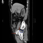
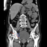
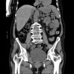
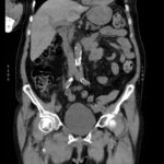
The non-contrast CT images show a minimally displaced comminuted fracture of the right acetabulum involving the acetabular roof, medial and anterior walls (red arrows), with associated obturator muscle hematoma (blue oval).
Discussion:
Acetabular fractures are quite rare. There are 37 pelvic fractures per 100,000 people in the United States annually, and only 10% of these involve the acetabulum. They occur more frequently in the elderly totaling an estimated 4,000 per year. High-energy trauma is the primary cause of acetabular fractures in younger individuals and these fractures are commonly associated with other fractures and pelvic ring disruptions. Fractures secondary to moderate or minimal trauma are increasingly of concern in patients of advanced age.1
Classification of acetabular fractures can be challenging. However, the approach can be simplified by remembering the three basic types of acetabular fractures (column, transverse, and wall) and their corresponding radiologic views. First, column fractures should be evaluated with coronally oriented CT images. This type of fracture demonstrates a coronal fracture line running caudad to craniad, essentially breaking the acetabulum into two halves: a front half and a back half. Secondly, transverse fractures should be evaluated by sagittally oriented CT images. By definition, a transverse fracture separates the acetabulum into superior and inferior halves with the fracture line extending from anterior to posterior; they will show a sagittally oriented fracture line at the roof of the acetabulum on axial CT. Lastly, wall fractures should be evaluated with axial CT images. This is because wall fractures have an obliquely oriented fracture line on axial CT images at the roof of the acetabulum, as opposed to the coronal and sagittal fracture lines described with column and transverse fractures, respectively.2 Fractures are organized using the Letournel Classification based on whether the fracture site lies in the anterior or posterior walls and columns of bone.
After diagnosis, early surgical intervention is critical in achieving good results.3 The majority of acetabular fractures are repaired by open reduction and internal fixation. Patients with significant osteopenia or communition benefit most from total hip arthroplasty. However, due to the complex nature of these fractures, there is potential for poor outcome regardless of the injury pattern due to contributing factors such as imperfect reduction, osteochondral defects in the acetabulum or femoral head, osteoarthritis, avascular necrosis of the femoral head, sciatic nerve injury and infection.4
Topics:
Acetabulum, acetabular fracture, hip fracture, orthopedics, ortho.
References:
- Mears DC, Velyvis JH, Chang CP.Displaced acetabular fractures managed operatively: indicators of outcome. Clin Orthop Relat Res. 2003;407:173-86.
- Lawrence DA, Menn K, Baumgaertner M, Haims AH. Acetabular fractures: anatomic and clinical considerations. AJR Am J Roentgenol. 2013;201(3):W425-W436. doi: 10.2214/AJR.12.10470
- Munshi N, Abbas A, Gulamhussein MA, Mehboob G, Qureshi RA. Functional outcome of the surgical management of acute acetabular fractures. Journal of Acute Disease. 2015:4(4):327-330. doi: 10.1302/0301-620X.98B5.36292
- Briffa N, Pearce AM, Hill M. Bircher. Outcomes of acetabular fracture fixation with ten years’ follow-up. J Bone Joint Surg Br. 2011;93(2):229-236. doi: 10.1302/0301-620X.93B2.24056


