Issue 5:1
Hemorrhagic Renal Cyst
DOI: https://doi.org/10.21980/J8C92VBedside renal ultrasound demonstrated a right renal cyst with echogenic debris consistent with a hemorrhagic cyst (red arrow). In addition, a computed tomography (CT) scan of the abdomen and pelvis revealed a 4mm non-obstructing right renal stone and bilateral renal cysts. The CT also confirmed the ultrasound finding of a right renal cyst with mild perinephric stranding possibly consistent with a hemorrhagic cyst.
Meckel’s Diverticulum Causing Small Bowel Intussusception in Third Trimester Pregnancy
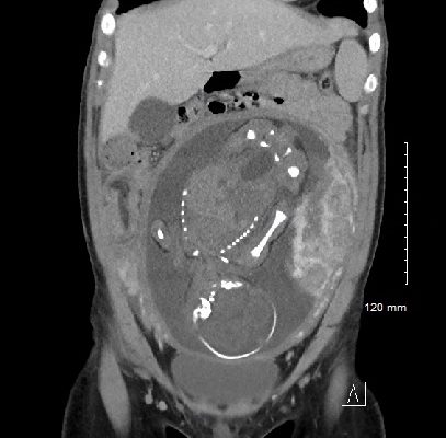
DOI: https://doi.org/10.21980/J87H19A CT scan was obtained which demonstrated distal small bowel intussusception with focal dilation suggestive of a small bowel obstruction in a pregnant female in her third trimester of pregnancy. The fetus can be seen in the uterus. The yellow arrow identifies the area of small bowel intussusception shown by telescoping intestines with associated bowel wall edema.
Bilateral Common Iliac Artery Aneurysm
DOI: https://doi.org/10.21980/J83S73A bedside ultrasound of the aorta was performed. The proximal, middle, and distal aorta appeared normal in caliber, as demonstrated by the images; however there seemed to be some enlargement at the bifurcation. The bifurcation into the iliac arteries, as highlighted by the yellow arrow, demonstrates a slightly enlarged iliac artery on the left. The aorta was followed below the bifurcation as it divided into the iliac arteries, as shown in the video clip. The ultrasound demonstrated a left iliac artery aneurysm measuring 5.99 cm, as highlighted by the orange circle. There were aneurysms to the bilateral common and internal iliac arteries.
Case Report: Acute Supraglottitis
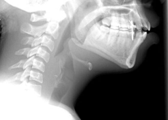
DOI: https://doi.org/10.21980/J8006VOn arrival, radiographs of the neck soft tissues were obtained, which showed a markedly enlarged epiglottic shadow (red arrow) concerning for epiglottitis. A computed tomography scan of the neck soft tissues with contrast was then obtained which revealed edematous mucosal thickening of the oropharynx (blue arrow) and supraglottic larynx (green arrow) including the epiglottis (purple arrow) concerning for acute infectious pharyngitis and supraglottic laryngitis with severe narrowing of the supraglottic laryngeal lumen, as well as associated extensive inflammation and edema of the superficial and deep left neck spaces. The patient’s white blood cell count was elevated to 25.7x10⁹/L with 87% neutrophils. Her rapid strep test was positive. Otolaryngology was consulted and performed a bedside flexible laryngoscopy which showed significant edema of the epiglottis (orange arrow), vocal cords (white arrow), and arytenoids (black arrow), left greater than right. Based on the findings and concern for impending respiratory failure, the patient received an awake fiberoptic intubation by anesthesia at the bedside.
Case Report of the Unusual Presentation of Stridor in an Elderly Patient Following a Cervical Fracture
DOI: https://doi.org/10.21980/J8V926The cervical CT was significant for a transverse fracture through the C4 vertebral body (see red arrow), lateral facet (green arrow), spinous process (blue arrow), and right lamina (purple arrow) as well as surrounding edema and retropharyngeal thickening (yellow line), best appreciated on sagittal view.
Henoch-Schönlein Purpura in the Adult
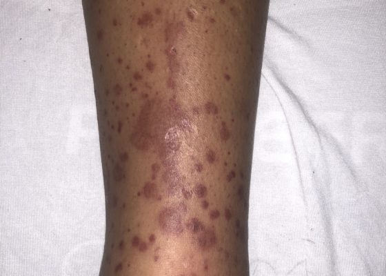
DOI: https://doi.org/10.21980/J8QH08The images show a raised, palpable, purpuric rash on the lower extremities, surrounded by a mild, 1+ non-pitting edema. Several of the lesions are exfoliated with serous discharge. There is no surrounding erythema, fluctuance, or lymphangitis to suggest cellulitis. There was no tenderness to palpation; however, pruritus was exacerbated on palpation.
Digital Nerve Block for the Reduction of a Proximal Phalanx Fracture of the Foot – a Case Report
DOI: https://doi.org/10.21980/J8KS8TPlain film of the right foot showed evidence of an oblique fracture of the body of the proximal 4th phalanx (image 2). No other acute traumatic injuries noted in the rest of the bones and joints of the right foot. After performing a digital block of the toe and reduction, repeat imaging showed evidence of successful reduction with anatomic alignment and redemonstration of the fracture line (image 3).
Case Report: Antifreeze Ingestion and Urine Fluorescence
DOI: https://doi.org/10.21980/J8G05TThe patient’s urine sample (right) was compared to a control (left) using a Wood’s lamp. It revealed light green fluorescence under ultraviolet light, which increased suspicion for ethylene glycol poisoning from antifreeze ingestion.
‹2
Page 2 of 2


