Renal and Splenic Infarcts
History of present illness:
A 61-year-old, diabetic, hypertensive woman presented to the emergency department in rapid atrial fibrillation with excruciating, non-tender, left upper quadrant and flank pain. She ran out of her anticoagulant a month before which she had been prescribed after a stroke earlier in the year.
Significant findings:
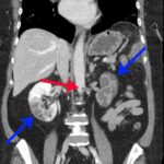
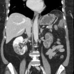
On the coronal sections of computed tomography (CT), bilateral renal infarctions (blue arrows) and several splenic infarctions (green arrows) are noted. Of particular interest, part of the clot totally occluding the left renal artery visibly extends into the aorta (red arrow). The vascular reconstruction image is remarkable for the absent left kidney, the unusual contour of the right kidney and the abnormal splenic blush.
Discussion:
Classic emergency medicine teaching dictates that when a patient with atrial fibrillation has abdominal pain “out of proportion” to the examination, one must consider mesenteric ischemia. Although the bowels clearly carry the highest embolic risk for abdominal viscera, other organs are at risk as well. This case illustrates a rare constellation of segmental splenic infarcts and bilateral renal infarction, with complete left renal artery occlusion stemming from multiple emboli.
Renal infarctions are difficult to predict because symptoms overlap with other potential etiologies such as abdominal aortic aneurysm, nephrolithiasis, mesenteric ischemia, appendicitis, and urinary tract infection.1 Patients with these infarctions typically have complaints such as flank or abdominal pain, nausea, vomiting, and sometimes fever.2 Splenic emboli produce similar nonspecific left-sided symptoms.The index of suspicion may be increased if the patient’s underlying disease state includes embolic risk factors such as atrial fibrillation, ischemic heart disease, infective endocarditis, cardiomyopathy, or valvular disease.3 The key to diagnosis in the emergency department is a CT scan with contrast, which has a sensitivity of over 95% in renal infarction cases.4
Due to the rarity of splenic and renal embolic infarctions, a standard treatment protocol has not been developed.2 Treatment may include anticoagulation, thrombolysis, surgical management, or interventional radiology.5 Our patient had the left renal artery clot extracted by the interventional radiologist followed by anticoagulation.
Topics:
Renal infarction, splenic infraction, embolic complications
References:
- Yousuf T, Ziffra J, Iqbal H, et al. Two cases of acute renal infarction in the setting of atrial fibrillation. Ochsner J. 2016;16(3):312–314.
- Bourgault M, Grimbert P, Verret C, et al. Acute renal infarction: a case series. Clin J Am Soc Nephrol. 2013;8(3):392-398. doi: 10.2215/CJN.05570612
- Bouassida K, Hmida W, Zairi A, et al. Bilateral renal infarction following atrial fibrillation and thromboembolism and presenting as acute abdominal pain: a case report. J Med Case Rep. 2012; 6:153. doi: 10.1186/1752-1947-6-153
- Lavoignet C, Le Borgne P, Ugé S, et al. Bilateral renal infarction after discontinuation of anticoagulant therapy. Nephrologie & therapeutique. 2016;12:234. doi:1016/j.nephro.2016.01.014
- Komolafe B, Dishmon D, Sultan W, Khouzam RN. Successful aspiration and rheolytic thrombectomy of a renal artery infarct and review of the current literature. Can J Cardiol. 2012;28(6):760-e1. doi: 10.1016/j.cjca.2012.06.020


