A Rare Cause of Pelvic Pain in a Teenage Girl
History of present illness:
A 14-year-old female presented with rectal pain, pelvic pressure, urinary hesitancy and difficulty defecating despite daily laxative use. She had a history of irregular periods and was currently menstruating. Her vital signs were normal. Her abdominal exam was unremarkable and the external genitourinary exam showed a visible vaginal introitus and no masses.
Significant findings:
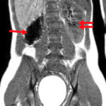
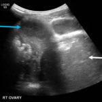
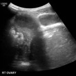
Due to pain out of proportion to her exam, an ultrasound of her pelvis was obtained and showed a blood-filled distended uterus, or hematometrocolpos (white arrow), with a 4.9 cm right ovarian cyst (blue arrow). A pelvic magnetic resonance imaging (MRI) then revealed an obstructed right hemi-vagina, normal left uterus and vagina and ipsilateral renal agenesis (red arrow) with normal left kidney (double arrow) consistent with obstructed hemivagina, ipsilateral renal agenesis (OHVIRA) syndrome. The patient underwent surgical repair with complete resolution of symptoms.
Discussion:
OHVIRA is rare syndrome that occurs due to a failure of lateral fusion of the Mullerian ducts.1 It affects an estimated 0.1%-3.8% of the population.2 The majority of these cases are associated with ipsilateral renal anomalies, but up to half can have contralateral anomalies.1,2 The most common presenting symptom is pelvic pain.1 On physical exam, patients may have a bulge in the vaginal wall which represents hematometrocolpos due to obstruction of outflow of menstrual blood. Transabdominal pelvic ultrasound or MRI are helpful modalities for diagnosis in adolescents.1,3 In one pediatric case series of OHVIRA, all 8 patients had a history of normal menses and presented with acute or chronic pelvic pain that began after menarche.2 All patients in this case series were initially misdiagnosed, due to the rarity of the disorder and the fact that most patients have a history of regular menses despite progressive pelvic pain. Definitive treatment requires surgical excision of the vaginal septum. Post-surgical prognosis is typically good;4-6 potential complications include endometriosis and increased risk for preterm labor or malpresentation.7
Topics:
Pelvic pain, obstructed hemivagina, renal agenesis, rectal pain, hematometrocolpos, hematocolpos.
References:
- Santos XM, Dietrich JE. Obstructed hemivagina with ipsilateral renal anomaly. J Pediatr Adolesc Gynecol. 2016; 29:7-10. https://doi.org/10.1016/j.jpag.2014.09.008.
- Zurawin RK, Dietrich JE, Heard MJ, Edwards CL. Didelphic uterus and obstructed hemivagina with renal agenesis: case report and review of the literature. J Pediatric Adolesc Gynecol.2004;17(2):137-141. doi: 10.1016/j.jpag.2004.01.016.
- Santos XM, Krishnamurthy R, Bercaw-Pratt JL, Dietrich JE. The utility of ultrasound and magnetic resonance imaging versus surgery for the characterization of Mullerian anomalies in the pediatric and adolescent population. J Pediatr Adolesc Gynecol. 2012;25(3):181-184. doi: 10.1016/j.jpag.2011.12.069.
- Heinonen, PK. Clinical implications of the didelphic uterus: long-term follow-up of 49 cases. European Journal of Obstetrics & Gynecology and Reproductive Biology. 2000;91(2): 183-190. doi: 10.1016/S0301-2115(99)00259-6.
- Smith NA, Laufer MR. Obstructed hemivagina and ipsilateral renal anomaly (OHVIRA) syndrome: management and follow-up. Fertility and Sterility. 2007;87(4):918-922.doi: 1016/j.fertnstert.2006.11.015.
- Rieger MM, Santos XM, Sangi-Haghpeykar H, Bercaw JL, Dietric JE. Laparoscopic outcomes for pelvic pathology in children and adolescents among patients presenting to the pediatric adolescent gynecology service. J Pediatr Adolesc Gynecol. 2015; 28(3):157-162. doi:1016/j.jpag.2014.06.008.
- Dietrich JE, Millar DM, Quint EH. Obstructive reproductive tract anomalies. J Pediatri Adolesc Gynecol.2014;27(6):396-402.doi: 1016/j.jpag.2014.09.001.


