Latest Articles
Acetaminophen Toxicity
DOI: https://doi.org/10.21980/J8435RAt the end of this practice oral board session, examinees will be able to: 1) demonstrate an ability to obtain a complete medical history in an oral boards structured interview format, 2) review appropriate laboratory tests and imaging to evaluate abdominal pain, 3) investigate a broad differential diagnosis for right upper quadrant abdominal pain, 4) recognize chronic acetaminophen toxicity, 5) initiate the appropriate treatment for chronic acetaminophen toxicity, 6) demonstrate effective communication with the patient, consultants, and the admitting team.
Journal Court: A Novel Approach to Incorporate Medicolegal Education into an Emergency Medicine Journal Club
DOI: https://doi.org/10.21980/J8093TBy the end of this exercise, participants should: 1) identify the four necessary elements for a malpractice claim, 2) understand the basic structure of medical malpractice litigation, and 3) critically analyze medical literature representing diverging viewpoints or conclusions.
A Cold Case: Myxedema Coma
DOI: https://doi.org/10.21980/J8VM0JAt the conclusion of the simulation, the learner is expected to: 1) Recognize the key features on history and examination of a patient presenting in myxedema coma and initiate the appropriate workup and treatment, 2) Describe clinical features and management for a patient with myxedema coma, 3) Develop a differential diagnosis for a critically ill patient with altered mental status, 4) Discuss the management of myxedema coma in the ED, including treatments, appropriate consultation, and disposition.
Drowning Complicated by Hypothermia
DOI: https://doi.org/10.21980/J8QS7PAt the conclusion of the simulation session, learners will be able to: 1) obtain a relevant focused history, including circumstances of drowning and/or cold exposure; 2) outline different clinical presentations of hypothermia, loosely correlated with core temperature readings; 3) discuss management of hypothermia, including passive external rewarming, active external rewarming, active internal rewarming, and extracorporeal blood rewarming; 4) discuss pathophysiology of drowning; 5) identify appropriate disposition of patients who present after drowning; and 6) identify appropriate disposition of hypothermic patients.
A Case Report of Right Atrial Thrombosis Complicated by Multiple Pulmonary Emboli: POCUS For the Win!
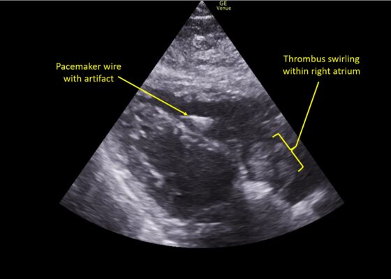
DOI: https://doi.org/10.21980/J8TM07Pulmonary POCUS was performed by the ED physician (GE Venue, C1-5-RS 5MHz curvilinear transducer), and lung examination was unremarkable with no pleural effusion, pneumothorax, or infiltrate. Subxiphoid views (GE Venue, 3Sc-RS 4MHz phased-array transducer) were obtained because this patient’s COPD with severe pulmonary hyperexpansion made parasternal and apical 4-chamber views suboptimal. A large thrombus can be seen within the right atrium (movie 1, images 1, 2). This has a serpiginous, rounded appearance and is mobile, appearing to swirl within the right atrium with intermittent extrusion through the tricuspid valve. A pacemaker wire is also visible within the right ventricle as a non-moving, hyperechoic, linear structure with posterior enhancement artifact. Pericardial effusion is not present.
Retropharyngeal Abscess in an Adult Patient Presenting with Neck Fullness and Dysphagia: A Case Report
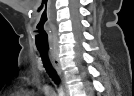
DOI: https://doi.org/10.21980/J8M36GContrast-enhanced CT soft tissue of the neck showed evidence of a prevertebral/retropharyngeal fluid collection, extending from the odontoid tip to the inferior C4 vertebral body margin, measuring 5.4 x 1.0 x 3.3 centimeters (cm) in size (yellow lines) without gross airway narrowing.
The Advantage of Using Video Laryngoscope in Puncture and Incisional Drainage of Peritonsillar Abscess: A Case Report
DOI: https://doi.org/10.21980/J8G935Incision of the peritonsillar abscess was performed with the assistance of the C-MAC video laryngoscope which provided a clear, illuminated, and unobstructed view of the incision site. Local anesthesia with 1% xylocaine was administered, and the abscess was incised with a scalpel and drained with a forceps.
A Case Report on an Elusive Incident of Erythema Multiforme
DOI: https://doi.org/10.21980/J8BM0WHer physical exam was notable for multiple scattered tense vesicles on an erythematous base along the left and right lower extremities and right upper extremity. The lesions were excoriated and in different stages of evolution. No oral, mucosal, or conjunctival lesions were found. Physical exam was otherwise unremarkable.
A Case of Painful Visual Loss – Managing Orbital Compartment Syndrome in the Emergency Department
DOI: https://doi.org/10.21980/J8N35DBy the end of this simulation, learners will be able to: 1) demonstrate the major components and a systematic approach to the emergency ophthalmologic examination, 2) develop a differential diagnosis of sight-threatening etiologies that could cause eye pain or vision loss, 3) demonstrate proficiency in performing potentially vision-saving procedures within the scope of EM practice.


