Respiratory
Pediatric Airway Team Based Learning
DOI: https://doi.org/10.21980/J8KH01This cTBL covers a variety of pediatric airway emergencies. Therefore, by the end of this cTBL, the learner will be able to: 1) List the signs and symptoms associated with airway foreign body obstructions. 2) State the appropriate management of upper and lower airway foreign bodies. 3) Discuss the symptoms, signs, and management of bacterial tracheitis. 4) Discuss a step-wise algorithm for emergency asthma treatment in the emergency department setting. 5) Identify the potential complications of tonsillectomy and the acute management of post-tonsillectomy hemorrhage.
Subcutaneous Emphysema After Chest Trauma
DOI: https://doi.org/10.21980/J8864NPlain film anteroposterior (AP) radiography of the chest shows left-sided subcutaneous emphysema (red arrow) with overlapping muscle striations of the pectoralis major (green arrow). After chest tube placement (blue arrow), AP chest radiography shows persistent left-sided subcutaneous emphysema (red arrow). CT of the chest shows pneumomediastinum (blue arrow), left apical pneumothorax (pink arrow), and subcutaneous emphysema (red arrow) at the level of T2. At the level of T6, rib fractures can be visualized on the CT (yellow arrow). At the level of T8, left sided pneumothorax is also seen (pink arrow) as the absence of lung tissue on CT.
Croup
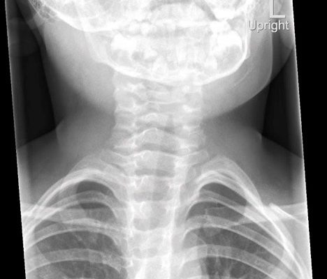
DOI: https://doi.org/10.21980/J8W05JThe anteroposterior X-ray reveals the classic steeple sign (blue outline) indicative of subglottic edema leading to tracheal narrowing, consistent with croup. The lateral x-ray shows narrowing of the subglottic region (green outline and arrows).
Bullous Emphysema
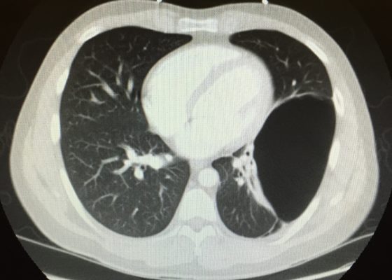
DOI: https://doi.org/10.21980/J8W62GThe upright chest X-ray shows a large lucent area in the left lower lung field without lung markings, with associated curvilinear opacities (yellow arrows) consistent with a large air-filled bulla. The bulla is large enough to compress adjacent lung tissue as shown by the visible pleural line (blue line). The discontinuity of the pleural line and presence of lung markings superiorly makes these findings more consistent with bulla than pneumothorax. The chest computed tomography (CT) confirmed a large left hemithorax bulla.
The Role of Chest X-Ray and Bedside Ultrasound in Diagnosing Pulmonary Bleb versus Pneumothorax
DOI: https://doi.org/10.21980/J8MP7QThe patient was evaluated with bedside ultrasound for concern of possible pneumothorax. Imaging of the left lung with M-mode demonstrated a “sea shore” sign showing a wavy pattern below the pleural line caused by lung sliding as well as “comet tail” artifact caused by from the deep pleura. However, there was no lung sliding on the right shown by a lack of “comet tail” artifact and a “bar code” sign where M-mode shows straight lines throughout the image, this is caused by lack of motion below the pleura. This lack of lung sliding is consistent with possible pneumothorax or bleb.
A two-view chest X-ray (CXR) revealed absent lung parenchyma in the right lung similar to a large pneumothorax (see red outline). Electronic medical record chart review revealed previous CXRs with similar findings. This patient was determined to have an acute COPD exacerbation with chronic blebs, but no pneumothorax.
Pediatric Foreign Body Aspiration
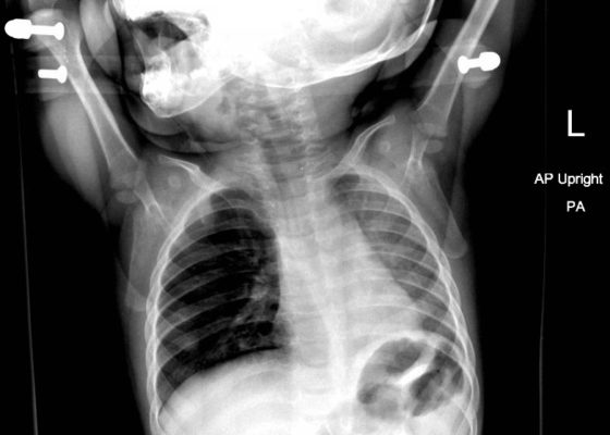
DOI: https://doi.org/10.21980/J8B648Chest radiograph showed increased radiolucency (red arrow) and flattening of the diaphragm on the right side (blue arrow) consistent with hyperinflation of the right lung, as well as left mediastinal shift (green arrow), indicating obstruction.
Spontaneous Pneumomediastinum: Hamman Syndrome
DOI: https://doi.org/10.21980/J8NS72The initial CT scans showed extraluminal gas surrounding the distal esophagus as it traversed the posterior mediastinum, concerning for possible distal esophageal perforation that prompted surgery and GI consultations. There was no evidence of a drainable collection or significant fat stranding. The image also showed an intraluminal stent traversing the gastric antrum and gastric pylorus with no indication of obstruction. Circumferential mural thickening of the gastric antrum and body were consistent with the patient’s history of gastric adenocarcinoma. The shotty perigastric lymph nodes with associated fat stranding, along the greater curvature of the distal gastric body suggested local regional nodal metastases and possible peritoneal carcinomatosis.
The thoracic CT scans showed extensive pneumomediastinum that tracked into the soft tissues of the neck, which given the history of vomiting also raised concern for esophageal perforation. There was still no evidence of mediastinal abscess or fat stranding. Additionally, a left subclavian vein port catheter, which terminates with tip at the cavoatrial junction of the superior vena cava can also be seen on the image.
Pediatric Pulmonary Abscess
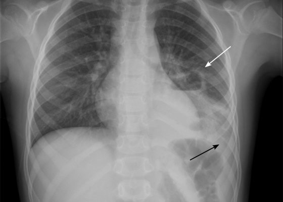
DOI: https://doi.org/10.21980/J83S6QUpright posterior-anterior plain chest films show a left lower lobe consolidation with an air-fluid level and a single septation consistent with a pulmonary abscess (white arrows). A small left pleural effusion was also present, seen as blunting of the left costophrenic angle and obscuration of the left hemidiaphragm (black arrows).
Pneumomediastinum After Cervical Stab Wound
DOI: https://doi.org/10.21980/J87P79Anteroposterior (AP) chest X-ray showed subcutaneous emphysema of the neck, surrounding the trachea (red arrows), right side greater than left, and a streak of gas adjacent to the aortic arch (white arrow). Computed tomography angiogram (CTA) of the neck showed air outside of the trachea, positive for pneumomediastinum (blue arrows).
Tricuspid Annular Plane Systolic Excursion (TAPSE) in a Patient with Pulmonary Emboli
DOI: https://doi.org/10.21980/J8M052Video 1 and Image 1 show a thrombus in the patient’s IVC. Video 2 and Images 2-3 demonstrate a positive TAPSE of less than 17mm (blue arrow length) with a significantly dilated RV, indicating abnormal excursion of the tricuspid annulus and right ventricular dysfunction.


