• Registration
  • Login
JETem
  • Home
  • About
    • Aim and Scope
    • Our Team
    • Editorial Board
    • FAQ
  • Issues
    • Current Issue
    • Ahead of Print
    • Past Issues
  • Visual EM
    • Latest Visual EM
    • Search Visual EM
    • Thumbnail Library
  • For Authors
    • Instructions for Authors
    • Submit to JETem
    • Photo Consent
    • Policies
      • Peer Review Policy
      • Copyright Policy
      • Editorial Policy, Ethics and Responsibilities
      • Conflicts of Interest & Informed Consent
      • Open Access Policy
  • For Reviewers
    • Instructions for JETem Reviewers
    • Interested in Being a JETem Reviewer?
  • Topic
    • Abdominal / Gastroenterology
    • Administration
    • Board Review
    • Cardiology / Vascular
    • Clinical Informatics, Telehealth and Technology
    • Dermatology
    • EMS
    • Endocrine
    • ENT
    • Faculty Development
    • Genitourinary
    • Geriatrics
    • Hematology / Oncology
    • Infectious Disease
    • Miscellaneous
    • Neurology
    • Ob / Gyn
    • Ophthalmology
    • Orthopedics
    • Pediatrics
    • Pharmacology
    • Procedures
    • Psychiatry
    • Renal / Electrolytes
    • Respiratory
    • Social Determinants of Health
    • Toxicology
    • Trauma
    • Ultrasound
    • Urology
    • Wellness
    • Wilderness
  • Modality
    • Curricula
    • Innovations
    • Lectures
    • Oral Boards
      • Structured Interview
      • Communication Case
    • Podcasts
    • Simulation
    • Small Group Learning
    • Team Based Learning
    • Visual EM
  • Contact Us

Ophthalmology

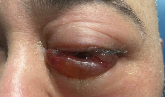
Inferior Rectus Abscess CT Coronal Unannotated. JETem 2025

A Case Report of Inferior Rectus Abscess

Luke Chi*, Adam Sauer, MD ^ and Danielle Matonis, MD^

DOI: https://doi.org/10.21980/J8J35G Issue 10:2 No ratings yet.
Non-contrast computed tomography (CT) imaging of the head in coronal, sagittal, and axial planes revealed a distinct 1.7 x 2.2 x 1.4 cm peripherally enhancing fluid collection within the left inferior orbit, involving the inferior rectus (yellow circle). This lesion resulted in restricted extraocular motility due to structural compression of the left globe. Laboratory results showed a mildly elevated white blood cell count of 11.5/mm3 and otherwise normal results including C-reactive protein (CRP) and erythrocyte sedimentation rate (ESR).
Visual EMCurrent IssueInfectious DiseaseOphthalmology

A Case of Painful Visual Loss – Managing Orbital Compartment Syndrome in the Emergency Department

Jessica Pelletier, DO*, Alexander Croft, MD*, Michael Pajor, MD*, Matthew Santos, MD^, Douglas Char, MD *, Marc Mendelsohn, MD, MPH*, and Ernesto Romo, MD* 

DOI: https://doi.org/10.21980/J8N35D Issue 9:4 No ratings yet.
By the end of this simulation, learners will be able to: 1) demonstrate the major components and a systematic approach to the emergency ophthalmologic examination, 2) develop a differential diagnosis of sight-threatening etiologies that could cause eye pain or vision loss, 3) demonstrate proficiency in performing potentially vision-saving procedures within the scope of EM practice.
OphthalmologySimulation
Internuclear Ophthalmoplegia. Eyes Right Unannotated. JETem 2024

The Clue is in the Eyes. A Case Report of Internuclear Ophthalmoplegia

Cooper Nickels, DO*, Christy Keyes, MD*, Caroline Astemborski, MD, MEHP* and Haley Fulton, BS^

DOI: https://doi.org/10.21980/J8DP9M Issue 9:1 No ratings yet.
There was no appreciable esotropia or exotropia noted on straight gaze (yellow arrows). On extraocular muscle examination, patient was noted to have a complete left medial rectus palsy consistent with a left internuclear ophthalmoplegia (red arrow). This was evidence by both eyes easily gazing left (green arrows); however, with rightward gaze, her left eye failed to gaze past midline (red arrow).
Visual EMNeurologyOphthalmology
CRAO Exam. JETem 2023

Sonographic Retrobulbar Spot Sign in Diagnosis of Central Retinal Artery Occlusion: A Case Report

Emiliya Usheva, MD*, Dustin Williams, MD^, Haley Musgrave, MD† and Scott Zhou, MD**

DOI: https://doi.org/10.21980/J8735P Issue 8:4 No ratings yet.
The bedside ocular ultrasound (B-scan) was significant for small, hyperechoic signal (white arrow) in the distal aspect of the optic nerve, concerning for embolus in the central retinal artery. Subsequent direct fundoscopic exam was significant for a pale macula with cherry red spot (black arrow), consistent with central retinal artery occlusion (CRAO).
OphthalmologyUltrasoundVisual EM
Mucormycosis. Photograph 2. JETem 2023

A Case Report of Invasive Mucormycosis in a COVID-19 Positive and Newly-Diagnosed Diabetic Patient

Konnor Davis, BS*, Roy Almog, MD^, Yuval Peleg, MD, MBA^ and Lindsey Spiegelman, MD, MBA^

DOI: https://doi.org/10.21980/J81M1G Issue 8:3 No ratings yet.
On physical exam, when the patient was asked to try and look to her right, the right eye failed to move laterally/abduct (blue arrow). Additionally, when asked to look straight ahead, the eye was slightly adducted (red arrow). There was a lack of motion of the right eye in abduction when the patient was asked to look to her right (yellow arrow).
Infectious DiseaseOphthalmologyVisual EM
Creative Commons images

Case Report of Herpes Zoster Ophthalmicus with Concurrent Parotitis

Serena Tally, BS*, Michelle Brown, DO* and Edmund Hsu, MD*

DOI: https://doi.org/10.21980/J8R93N Issue 8:2 No ratings yet.
The presence of soft tissue stranding about the parotid gland suggested an underlying inflammatory or infectious process of the parotid gland. Cellulitis was considered as a possible diagnosis as well, given the presence of soft tissue stranding in the dermis that is adjacent to the parotid gland. Fortunately, no enhancement was seen in local muscles, fascia, or bones to suggest a myositis, fasciitis, or osteomyelitis. By using the anatomy of the patient and understanding the changes that occur on CT when inflammation is present, the appropriate depth and location of infection can be made, allowing for appropriate treatment regimens.
Visual EMInfectious DiseaseOphthalmology

Use of An Ophthalmology Tutorial to Improve Resident Comfort with the Emergency Eye Exam

Jessica Pelletier, DO*, John Facciani, MD^, Francesca Gines, COA^ and Damon Kuehl, MD*

DOI: https://doi.org/10.21980/J86H0M Issue 7:4 No ratings yet.
By the end of this small group didactic, learners will be able to: 1) demonstrate ability to focus on the various components of the slit lamp exam 2) demonstrate understanding of a systematic approach to the eye exam 3) demonstrate appropriate use of the Diaton, iCare, and Tonopen tonometers.
OphthalmologySmall Group Learning
Creative Commons images

Vitreous Hemorrhage Case Report

Mary Rometti, MD*, Laryssa Patti, MD* and Christopher Bryczkowski, MD*

DOI: https://doi.org/10.21980/J88D3B Issue 7:3 No ratings yet.
Point of care ultrasound (POCUS) revealed hyperechoic material in the vitreous consistent with a vitreous hemorrhage. On the ultrasound images, there is visible hyperechoic debris seen floating in the vitreous as the patient moves his eye. Since the vitreous is typically anechoic (black) in color on ultrasound, turning up the gain on the ultrasound machine makes these findings easier to see and often highlights abnormalities, such as this hemorrhage (see annotated still).
OphthalmologyUltrasoundVisual EM
Creative Commons images

Case Report of a Man with Right Eye Pain and Double Vision

Nicolas Kahl, MD* and Maria Pelucio, MD*

DOI: https://doi.org/10.21980/J8KW7G Issue 7:1 No ratings yet.
ABSTRACT: A 39-year-old previously healthy male presented with three days of right eye pressure and one day of binocular diplopia.  He denied history of trauma, headache, or other neurological complaints.  He had normal visual acuity, normal intraocular pressure, intact convergence, and no afferent pupillary defect. His neurologic examination was non-focal except for an inability to adduct the right eye past midline
OphthalmologyNeurologyVisual EM

Build Your Own Eye: A Method for Teaching Ocular Anatomy and Pathophysiology

Alaina Brinley Rajagopal, MD, PhD*, Mark Joseph Slader, BS* and Megan Boysen Osborn, MD, MHPE*

DOI: https://doi.org/10.21980/J8GS8W Issue 5:3 No ratings yet.
By the end of this session the participant will be able to: 1) Describe basic anatomy of the eye, 2) Build a basic model of the eye, 3) Identify which diseases are associated with which parts of the eye, 4) Identify the pathophysiology behind diseases of the eye, 5) Name correct treatment for each disease.
OphthalmologyTeam Based Learning (TBL)
1›»
Page 1 of 3

JETem is an online, open access, peer-reviewed, journal-repository for EM educators. We are PMC Indexed.

Most Viewed

  • The Silent Saboteur: Teaching the Clinical Implications of Occult Hypoxemia & Social Determinants of Health via a Pulmonary Embolism Case
  • Diabetic Ketoacidosis and Necrotizing Soft Tissue Infection
  • My Broken Heart
  • Stabilization of Cardiogenic Shock for Critical Care Transport, a Simulation
  • Innovative Ultrasound-Guided Erector Spinae Plane Nerve Block Model for Training Emergency Medicine Physicians

Visit Our Collaborators

Creative Commons Licence
This work is licensed under a Creative Commons Attribution 4.0 International License.

About

Education

Learners should benefit from active learning. JETem accepts submissions of team-based learning, small group learning, simulation, podcasts, lectures, innovations, curricula, question sets, and visualEM.

Scholarship

We believe educators should advance through the scholarship of their educational work. JETem gives educators the opportunity to publish scholarly academic work so that it may be widely distributed, thereby increasing the significance of their results.

Links

  • Home
  • Aim and Scope
  • Current Issue
  • For Reviewers
  • Instructions for Authors
  • Contact Us

Newsletter

Sign up to receive updates from JETem regarding newly published issues and findings.

Copyright Creative Commons Attribution 4.0 International