• Registration
  • Login
JETem
  • Home
  • About
    • Aim and Scope
    • Our Team
    • Editorial Board
    • FAQ
  • Issues
    • Current Issue
    • Ahead of Print
    • Past Issues
  • Visual EM
    • Latest Visual EM
    • Search Visual EM
    • Thumbnail Library
  • For Authors
    • Instructions for Authors
    • Submit to JETem
    • Photo Consent
    • Policies
      • Peer Review Policy
      • Copyright Policy
      • Editorial Policy, Ethics and Responsibilities
      • Conflicts of Interest & Informed Consent
      • Open Access Policy
  • For Reviewers
    • Instructions for JETem Reviewers
    • Interested in Being a JETem Reviewer?
  • Topic
    • Abdominal / Gastroenterology
    • Administration
    • Board Review
    • Cardiology / Vascular
    • Clinical Informatics, Telehealth and Technology
    • Dermatology
    • EMS
    • Endocrine
    • ENT
    • Faculty Development
    • Genitourinary
    • Geriatrics
    • Hematology / Oncology
    • Infectious Disease
    • Miscellaneous
    • Neurology
    • Ob / Gyn
    • Ophthalmology
    • Orthopedics
    • Pediatrics
    • Pharmacology
    • Procedures
    • Psychiatry
    • Renal / Electrolytes
    • Respiratory
    • Social Determinants of Health
    • Toxicology
    • Trauma
    • Ultrasound
    • Urology
    • Wellness
    • Wilderness
  • Modality
    • Curricula
    • Innovations
    • Lectures
    • Certifying Exam Practice Cases
      • Clinical Care Cases
      • Communication & Procedural Cases
      • Traditional Oral Boards
    • Podcasts
    • Simulation
    • Small Group Learning
    • Team Based Learning
    • Visual EM
  • Contact Us

Endocrine

Nonketotic Hyperglycemia Hemichorea. MRI Unnnotated. JETem 2025

Case Report of a Patient Presenting with Nonketotic Hyperglycemia Hemichorea

Jay Patel, DO*, Kayla Pena, MD*, Joshua Bucher, MD* and Amanda Esposito, MD*

DOI: https://doi.org/10.21980/J8.52115Issue 10:4[mrp_rating_result]
Laboratory tests indicated elevated blood glucose levels (198 mg/dL) with no urinary ketones, anion gap of 12, thyroid stimulating hormone (TSH) of 12 UIU/ml, and an increased glycated hemoglobin (HbA1c) of 14.9%. After initial stroke evaluation with neurology, imaging studies, including computed tomography (CT)/CT angiography (CTA) of the brain and neck, were unremarkable, ruling out structural lesions or acute stroke. Neurology recommended an MRI which showed T1 shortening within the left basal ganglia involving both the caudate nucleus and the lentiform nucleus. T1 shortening indicates changes in the tissue composition or structure that alters how the tissue responds to the MRI pulse, giving the tissue a brighter appearance on MRI (see white arrow).
Visual EMCurrent IssueEndocrineNeurology

Diabetic Ketoacidosis and Necrotizing Soft Tissue Infection

Matthew Henschel, DO* and Stephanie Songey, DO^

DOI: https://doi.org/10.21980/J89M0K Issue 10:2[mrp_rating_result]
At the end of this oral board session, examinees will: 1) Demonstrate the ability to obtain a complete medical history and physical exam. 2) Identify and appropriately treat DKA. 3) Identify, treat, and make appropriate consults for NSTI. 4) Demonstrate effective communication of the treatment plan with the patient.
EndocrineInfectious DiseaseOral Boards

A Cold Case: Myxedema Coma

Andrew M Namespetra, MD*, Matthew J Petruso, DO* and Andrew M Bazakis, MD*

DOI: https://doi.org/10.21980/J8VM0J Issue 10:1[mrp_rating_result]
At the conclusion of the simulation, the learner is expected to: 1) Recognize the key features on history and examination of a patient presenting in myxedema coma and initiate the appropriate workup and treatment, 2) Describe clinical features and management for a patient with myxedema coma, 3) Develop a differential diagnosis for a critically ill patient with altered mental status, 4) Discuss the management of myxedema coma in the ED, including treatments, appropriate consultation, and disposition.
EndocrineSimulation

Adolescent with Diabetic Ketoacidosis, Hypothermia and Pneumomediastinum

Steven Millner, MD* and Courtney Devlin, MD* 

DOI: https://doi.org/10.21980/J8FP8J Issue 8:4[mrp_rating_result]
By the end of the simulation, learners will be able to: 1) develop a differential diagnosis for an adolescent who presents obtunded with shortness of breath; 2) discuss the management of diabetic ketoacidosis; 3) discuss management of hypothermia in a pediatric patient; 4) discuss appropriate ventilator settings in a patient with diabetic ketoacidosis; and 5) demonstrate interpersonal communication with family, nursing, and consultants during high stress situations.
EndocrinePediatricsProceduresRespiratorySimulation
Creative Commons images

A Case Report of Glycogenic Hepatopathy

Dane Brown, MD* and Theresa Mead, DO, RDMS*

DOI: https://doi.org/10.21980/J8SQ0Z Issue 6:3[mrp_rating_result]
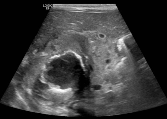
The ultrasound images reveal hepatomegaly and an increased echogenicity of the liver parenchyma that is diffuse. The increased echogenicity can be best appreciated by a comparison to surrounding structures. It is important to note that the increased echogenicity is non-focal and consistent throughout the entire liver in multiple views. These findings can be consistent with nonalcoholic steatohepatitis as well as glycogenic hepatopathy.
EndocrineVisual EM
Creative Commons images

A Case Report of a Large Goiter Resulting in Tracheal Deviation

Thomas Powell, MD* and Geremiha Emerson, MD*

DOI: https://doi.org/10.21980/J80645 Issue 6:3[mrp_rating_result]
In the image, one can see significant tracheal deviation around the right side of the mass (black arrows). This degree of deviation would make ventilation in a paralyzed patient extremely difficult, if not impossible.
EndocrineVisual EM
Creative Commons images

A Case Report of Neonatal Vomiting due to Adrenal Hemorrhage, Abscess and Pseudohypoaldosteronism

Raymen Rammy Assaf, MD, MPH* and Mary Jane Piroutek, MD^

DOI: https://doi.org/10.21980/J8QQ0B Issue 6:3[mrp_rating_result]
An ultrasound (US) of the abdomen was obtained to evaluate for possible pyloric stenosis (see US transverse/dopper imaging). While imaging showed a normal pyloric channel, it revealed an unexpected finding: a complex cystic mass arising from the right adrenal gland (yellow outline), measuring 5.8 by 4.0 by 6.4 cm with calcifications peripherally and mass effect on the kidney without evidence of vascular flow (blue arrow). Computed tomography (CT) of the abdomen/pelvis with IV contrast was subsequently obtained and measured the lesion as 2.8 by 4.6 by 4 cm without evidence of additional masses, lymphadenopathy or left adrenal gland abnormality (see CT axial, coronal, and sagittal imaging).
EndocrineVisual EM

Thyroid Storm

Kathryn Ritter, MD* and Carmen Wolfe, MD*

DOI: https://doi.org/10.21980/J8RW71 Issue 5:4[mrp_rating_result]
At the end of this practice oral boards case, the learner will: 1) assess a patient with altered mental status in an oral boards format; 2) review appropriate laboratory testing and diagnostic imaging; 3) identify signs and symptoms of thyroid storm and 4) review appropriate pharmacologic therapies with the proper sequence and timing.
EndocrineOral Boards
Creative Commons images

Case Report of Spontaneous Thyroid Hemorrhage Following LMA Insertion

Gregory Podolej, MD* and Gary Bhagat, MD*

DOI: https://doi.org/10.21980/J8XP8W Issue 5:3[mrp_rating_result]
Two photographs of patients neck, both showcasing no obvious erythema, bruising, or swelling which is noteworthy because there is potential for airway compromise but there was nothing visible to indicate that on exam. CTA of neck showing thyroid nodule and potential thyroid hemorrhage (outlined in orange) on the left without evidence of airway compromise at the time of CT scan. Official read by attending radiologist states there is a “heterogeneous left thyroid nodule measuring 3 cm. Findings are suggestive of multinodular goiter with possible acute hemorrhage. Adjacent tract of soft tissue stranding in the anterior left neck with mild adjacent fascial thickening. This could represent small amount of hemorrhage or could be inflammatory.”
EndocrineVisual EM
Creative Commons images

The Elusive Pheo: A Case Report of Pheochromocytoma in the Emergency Department

Jason Cody Pickett, MD*  and Jonah Gunalda, MD*

DOI: https://doi.org/10.21980/J8KW63 Issue 4:4[mrp_rating_result]
ED work-up: BMP within normal limits (WNL), white blood cell (WBC) 27.4, ECG showed sinus tachycardia, nonspecific ST segment abnormalities, BNP and troponin were within normal limits, HR 146 beats/minute, CT abdomen/pelvis showed a 10-cm-heterogenous enhancing left adrenal mass (anterior-posterior view [shown in red], lateral view [shown in blue]).
EndocrineVisual EM
1›
Page 1 of 2

JETem is an online, open access, peer-reviewed, journal-repository for EM educators. We are PMC Indexed.

Most Viewed

  • Novel Asynchronous Emergency Medicine Sub-Internship Curriculum Utilizing Free Open Access Medical Education (FOAM)
  • A Low-Cost Task Trainer Constructed from Silicone Nipple Covers
  • Pizza and Paintballs: A Cost-Effective Model for Incision and Drainage Simulation Training
  • Abdominal Pain and Vaginal Discharge: An Eye-Opening Simulation Case about Human Trafficking
  • The EMazing Race: A Novel Gamified Board and Clinical Practice Review for Emergency Medicine Residents

Visit Our Collaborators

Creative Commons Licence
This work is licensed under a Creative Commons Attribution 4.0 International License.

About

Education

Learners should benefit from active learning. JETem accepts submissions of team-based learning, small group learning, simulation, podcasts, lectures, innovations, curricula, question sets, and visualEM.

Scholarship

We believe educators should advance through the scholarship of their educational work. JETem gives educators the opportunity to publish scholarly academic work so that it may be widely distributed, thereby increasing the significance of their results.

Links

  • Home
  • Aim and Scope
  • Current Issue
  • For Reviewers
  • Instructions for Authors
  • Contact Us

Newsletter

Sign up to receive updates from JETem regarding newly published issues and findings.

Copyright Creative Commons Attribution 4.0 International