• Registration
  • Login
JETem
  • Home
  • About
    • Aim and Scope
    • Our Team
    • Editorial Board
    • FAQ
  • Issues
    • Current Issue
    • Ahead of Print
    • Past Issues
  • Visual EM
    • Latest Visual EM
    • Search Visual EM
    • Thumbnail Library
  • For Authors
    • Instructions for Authors
    • Submit to JETem
    • Photo Consent
    • Policies
      • Peer Review Policy
      • Copyright Policy
      • Editorial Policy, Ethics and Responsibilities
      • Conflicts of Interest & Informed Consent
      • Open Access Policy
  • For Reviewers
    • Instructions for JETem Reviewers
    • Interested in Being a JETem Reviewer?
  • Topic
    • Abdominal / Gastroenterology
    • Administration
    • Board Review
    • Cardiology / Vascular
    • Clinical Informatics, Telehealth and Technology
    • Dermatology
    • EMS
    • Endocrine
    • ENT
    • Faculty Development
    • Genitourinary
    • Geriatrics
    • Hematology / Oncology
    • Infectious Disease
    • Miscellaneous
    • Neurology
    • Ob / Gyn
    • Ophthalmology
    • Orthopedics
    • Pediatrics
    • Pharmacology
    • Procedures
    • Psychiatry
    • Renal / Electrolytes
    • Respiratory
    • Social Determinants of Health
    • Toxicology
    • Trauma
    • Ultrasound
    • Urology
    • Wellness
    • Wilderness
  • Modality
    • Curricula
    • Innovations
    • Lectures
    • Certifying Exam Practice Cases
      • Clinical Care Cases
      • Communication & Procedural Cases
      • Traditional Oral Boards
    • Podcasts
    • Simulation
    • Small Group Learning
    • Team Based Learning
    • Visual EM
  • Contact Us

Abdominal/Gastroenterology

Creative Commons images

Wandering Spleen

Jeffrey Nafash, MD, MPH* and Uchechi Azubuine, MD*

DOI: https://doi.org/10.21980/J8PS7C Issue 4:3 No ratings yet.
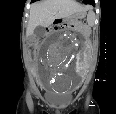
History of present illness: A 7-month-old boy presented for evaluation of an abdominal mass. Two weeks prior, the patient had a fever followed by 24 hours of vomiting and abdominal pain, which self-resolved. His pediatrician noted the mass on exam and referred child to the emergency department (ED). His abdominal exam was significant for a hard mass palpated to the
Abdominal/GastroenterologyVisual EM
Creative Commons images

Acute Pancreatitis

Ronald Goubert, BS*, Jonathan Peña, MD*, Alisa Wray, MD, MAEd* and Eleanor Chu, MD^

DOI: https://doi.org/10.21980/J88W5XIssue 4:2 No ratings yet.
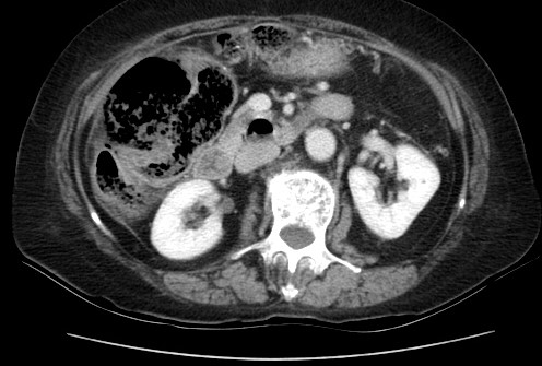
Computed tomography of the abdomen and pelvis with contrast show edema of the pancreas (red outline) and duodenum (yellow arrow) with peripancreatic inflammation, fluid and fat stranding (blue highlight). The distal pancreatic tail was noted to appear normal (green arrow). There was no organized drainable fluid collection, and no parenchymal hypo-enhancement. These findings are consistent with moderate severity acute interstitial pancreatitis.
Abdominal/GastroenterologyVisual EM
Creative Commons images

Renal and Splenic Infarcts

Niketu Patel, MPH* and Loice Swisher, MD*

DOI: https://doi.org/10.21980/J8804KIssue 4:2 No ratings yet.
On the coronal sections of computed tomography (CT), bilateral renal infarctions (blue arrows) and several splenic infarctions (green arrows) are noted. Of particular interest, part of the clot totally occluding the left renal artery visibly extends into the aorta (red arrow). The vascular reconstruction image is remarkable for the absent left kidney, the unusual contour of the right kidney and the abnormal splenic blush.
Visual EMAbdominal/Gastroenterology
Creative Commons images

Sigmoid Diverticulitis Complicated by Colovesical Fistula Presenting with Pneumaturia

Faraz Khan, BS*, Justin Yanuck, MD* and C Eric McCoy, MD, MPH*

DOI: https://doi.org/10.21980/J80G9TIssue 4:2 No ratings yet.
A CT scan of his abdomen/pelvis shows acute sigmoid colonic diverticulitis with adjacent extraluminal collection containing gas (axial view, white arrow) consistent with perforation, along with abutment of the urinary bladder with intraluminal bladder gas (sagittal and coronal views, white arrowheads) suggesting colovesical fistula.
Abdominal/GastroenterologyVisual EM
Creative Commons images

Brief Review of Intussusception Diagnosis and Management

Claire Thomas, MD*, Wirachin Hoonpongsimanont, MD*, Sara Paradise, MD* and Mohammad Helmy, MD^

DOI: https://doi.org/10.21980/J81P7FIssue 3:4 No ratings yet.
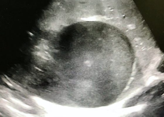
The patient’s abdominal ultrasound revealed intussusception in the right upper abdominal quadrant. The transverse ultrasound view showed a “doughnut sign” (dashed yellow line), telescoping bowel (yellow arrow), and invaginated hyperechoic mesenteric fat with crescent configuration (dashed orange line).  The sagittal ultrasound view demonstrated the intussusception formed by the outer recipient bowel loop (yellow arrows), invaginated hyperechoic mesenteric fat (orange asterisks), and telescoping bowel centrally (red arrow).
Abdominal/GastroenterologyPediatricsVisual EM
Creative Commons images

Realistic and Inexpensive Ultrasound Guided Paracentesis Simulator Using Pork Belly with Skin

Jonathan Kei, MD, MPH* and Donald P Mebust, MD*

DOI: https://doi.org/10.21980/J8NK9RIssue 3:3 No ratings yet.
By the end of this instructional session learners should be able to: 1) Discuss the indications, contraindications, and complications associated with abdominal paracentesis; and 2) competently perform an ultrasound-guided abdominal paracentesis on a simulator and remove fluid.
Abdominal/GastroenterologyInnovationsProcedures
Creative Commons images

Recurrent Sigmoid Volvulus in a Young Female

Ahmed Farhat, BS* and Robert Rowe, MD*

DOI: https://doi.org/10.21980/J8GW5SIssue 3:3 No ratings yet.
Computed tomography (CT) of the abdomen and pelvis was obtained revealing a colonic volvulus in the left mid to upper abdomen (blue arrow) involving the distal transverse colon and descending colon, with gaseous colonic distention to 8.5 cm (red arrow). The characteristic “whirl pattern” is also present (yellow arrow). These findings are suggestive of a high-grade colonic obstruction. It was without evidence of pneumoperitoneum, pneumatosis, or drainable collection. Of note, a 3.6 cm dermoid tumor is also observable in the left adnexa (green arrow).
Abdominal/GastroenterologyVisual EM
Creative Commons images

Acute Dysphagia in a 25-Year-Old Male

Michael Ullo, MD*, Robert Joshua Dym, MD^ and Jill Ripper, MD*

DOI: https://doi.org/10.21980/J83P8FIssue 3:3 No ratings yet.
After an unremarkable chest radiograph was obtained, a computed tomography (CT) scan of the chest was obtained due to possible co-ingestion of bones to rule out perforation. The CT scan demonstrated focal distention of the mid-esophagus due to an impacted food bolus (white arrow). An aberrant right subclavian artery (yellow arrow) was located just distal to the impaction site with partial compression of the esophagus (red arrow).
Abdominal/GastroenterologyVisual EM
Creative Commons images

Woman Swallows a “Handful of Pills”

Sarah E Mott, MD*, Michael Paddock, DO, MS*^ and Jessie Nelson, MD*^

DOI: https://doi.org/10.21980/J8V64XIssue 3:3 No ratings yet.
Soft tissue lateral X-ray of neck was performed. The lateral soft tissue X-ray of the neck showed a metallic foreign body at the level cricoid.
Abdominal/GastroenterologyVisual EM
Creative Commons images

Incidental Hiatal Hernia on Chest X-ray

Ahmed Farhat, BS* and Daryn Towle, MD*

DOI: https://doi.org/10.21980/J8KP8SIssue 3:3 No ratings yet.
The two-view chest X-ray shows mild opacification of the bilateral lower lobes concerning for pneumonia (red arrows). Incidental retrocardiac opacity with air-fluid level consistent with large hiatal hernia is also observed (green arrow).
Abdominal/GastroenterologyVisual EM
«‹4›»
Page 4 of 7

JETem is an online, open access, peer-reviewed, journal-repository for EM educators. We are PMC Indexed.

Most Viewed

  • A Longitudinal, Practical Curriculum for Faculty Development as New Coaches in Graduate Medical Education
  • Cognitive Errors and Debiasing
  • Critical Care Transport: Blunt Polytrauma in Pregnancy
  • Beta-Blocker Toxicity
  • A Comprehensive and Modality Diverse Cervical Spine and Back Musculoskeletal Physical Exam Curriculum for Medical Students

Visit Our Collaborators

Creative Commons Licence
This work is licensed under a Creative Commons Attribution 4.0 International License.

About

Education

Learners should benefit from active learning. JETem accepts submissions of team-based learning, small group learning, simulation, podcasts, lectures, innovations, curricula, question sets, and visualEM.

Scholarship

We believe educators should advance through the scholarship of their educational work. JETem gives educators the opportunity to publish scholarly academic work so that it may be widely distributed, thereby increasing the significance of their results.

Links

  • Home
  • Aim and Scope
  • Current Issue
  • For Reviewers
  • Instructions for Authors
  • Contact Us

Newsletter

Sign up to receive updates from JETem regarding newly published issues and findings.

Copyright Creative Commons Attribution 4.0 International