Recurrent Sigmoid Volvulus in a Young Female
History of present illness:
A 31-year-old female with history of sigmoid volvulus status post sigmoidectomy 2 years prior presented to the emergency department with 1-week history of diffuse, crampy abdominal pain. In the 24 hours prior to her arrival, patient stated that her abdominal pain acutely worsened and was accompanied with decreased stool output and non-bloody, non-bilious emesis. She denied any fever, chills, weight loss, chest pain, or shortness of breath.
Significant findings:
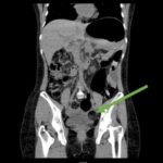
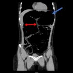
Computed tomography (CT) of the abdomen and pelvis was obtained revealing a colonic volvulus in the left mid to upper abdomen (blue arrow) involving the distal transverse colon and descending colon, with gaseous colonic distention to 8.5 cm (red arrow). The characteristic “whirl pattern” is also present (yellow arrow). These findings are suggestive of a high-grade colonic obstruction. It was without evidence of pneumoperitoneum, pneumatosis, or drainable collection. Of note, a 3.6 cm dermoid tumor is also observable in the left adnexa (green arrow).
Discussion:
A colonic volvulus refers to the twisting of a portion of the colon, most often the sigmoid, in a manner leading to obstruction and potential ischemia and gangrene.1 Colonic volvulus is responsible for approximately 15% of all large bowel obstructions in the United States.1 The mortality associated with this condition is less than 10% in patients who have not developed gangrene, but can be as high as 60% if gangrene is present.2 A sigmoid volvulus occurs when the sigmoid colon is elongated, leading to a redundant loop which rotates around its mesocolon.3 This condition is most common in the elderly with a mean age of 70 years at presentation.4 Institutionalized patients with neuropsychiatric disorders and patients in nursing homes are at the highest risk as these populations experience prolonged recumbency and chronic constipation, which are both highly associated with sigmoid volvulus development.1
Patients with sigmoid volvulus present with slow onset and progressive abdominal pain, nausea, abdominal distention, and constipation. Vomiting is also common but normally occurs several days after the initial onset of pain.5 Computed tomography is the preferred method of diagnosis with a sensitivity of 71%.6 Diagnostic findings include a “whirl pattern,” which is caused by the twisting of the sigmoid colon around its mesocolon, and a “bird-beak” appearance of the afferent and efferent colonic segments.7 Treatment of a sigmoid volvulus begins with a flexible sigmoidoscopy for decompression and detorsion, followed by definitive surgery to prevent recurrence.4 Without definitive surgery, recurrence rates of sigmoid volvulus has been reported as high as 90%.1 In this case, gastroenterology was consulted and patient was taken to the endoscopy suite for decompression and detorsion.
Topics:
Sigmoid volvulus, large bowel obstruction, colonic volvulus.
References:
- Gingold D, Murrell Z. Management of colonic volvulus. Clin Colon Rectal Surg. 2012;25(4):236-244. doi:10.1055/s-0032-1329535
- Mangiante EC, Croce MA, Fabian TC. Sigmoid volvulus. A four-decade experience. The American Surgeon.1989;55(1):41-44.
- Osiro SB, Cunningham D, Shoja MM, Tubbs RS, Gielecki J, Loukas M. The twisted colon: a review of sigmoid volvulus. Am Surg.2012;78(3):271-279.
- Halabi WJ, Jafari MD, Kang CY, Nguyen VQ, Carmichael JC, Mills S, et al. Colonic Volvulus in the United States: trends, outcomes, and predictors of mortality. Ann Surg.2014;259(2):293-301. doi: 10.1097/SLA.0b013e31828c88ac
- Atamanalp SS. Sigmoid volvulus: diagnosis in 938 patients over 45.5 years. Tech Coloproctol.2013;17(4):419-424. doi: 10.1007/s10151-012-0953-z
- Levsky JM, Den EI, DuBrow RA, Wolf EL, Rozenblit AM. CT findings of sigmoid volvulus. AJR Am J Roentgenol.2010;194(1):136-143. doi: 10.2214/AJR.09.2580
- Catalano O. Computed tomographic image of sigmoid volvulus. Abdom Imaging. 1996; 21:314-317. doi: 10.1007/s002619900


