Scaphoid Fracture
History of present illness:
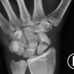
A 25-year-old, right-handed male presented to the emergency department with left wrist pain after falling from a skateboard onto an outstretched hand two-weeks prior. He otherwise had no additional concerns, including no complaints of weakness or loss of sensation. On physical exam, there was tenderness to palpation within the anatomical snuff box. The neurovascular exam was intact. Plain films of the left wrist and hand were obtained.
Significant findings:
The anteroposterior (AP) plain film of this patient demonstrates a full thickness fracture through the middle third of the scaphoid (red arrow), with some apparent displacement (yellow lines) and subtle angulation of the fracture fragments (blue line).
Discussion:
The scaphoid bone is the most commonly fractured carpal bone accounting for 70%-80% of carpal fractures.1 Classically, it is sustained following a fall onto an outstretched hand (FOOSH). Patients should be evaluated for tenderness with palpation over the anatomical snuffbox, which has a sensitivity of 100% and specificity of 40%.2 Plain films are the initial diagnostic modality of choice and have a sensitivity of 70%, but are commonly falsely negative in the first two to six weeks of injury (false negative of 20%).3 The Mayo classification organizes scaphoid fractures as involving the proximal, mid, and distal portions of the scaphoid bone with mid-fractures being the most common.3
The proximal scaphoid is highly susceptible to vascular compromise because it depends on retrograde blood flow from the radial artery. Therefore, disruption can lead to serious sequelae including osteonecrosis, arthrosis, and functional impairment. Thus, a low threshold should be maintained for neurovascular evaluation and surgical referral. Patients with non-displaced scaphoid fractures should be placed in a thumb spica splint.3 Patients with even suspected scaphoid fractures should be placed in a thumb spica splint and re-evaluated in two weeks with repeat plain films.4 Further evaluation with MRI or bone scan should be considered if repeat plain films are negative and clinical suspicion remains high.3 In conclusion, scaphoid fractures can be difficult to identify; therefore, a high level of suspicion and low threshold for further imaging or intervention is warranted.
Topics:
Scaphoid, fracture, orthopedics, FOOSH injury.
References:
- Pines JM, Everett WW. Evidence-Based Emergency Care, Diagnostic Testing and Clinical Decision Rules. Hoboken NJ: Wiley-Blackwell, BMJ Books; 2011.
- Phillips TG, Reibach AM, Slomiany WP. Diagnosis and management of scaphoid fractures. Am Fam Physician. 2004;70(5): 879-884.
- Rhemrev SJ, Ootes D, Beeres, FJ, Meylaerts SA, Schipper IB. Current methods of diagnosis and treatment of scaphoid fractures. Int J Emerg Med. 2011;4:4. doi: 10.1186/1865-1380-4-4
- Steinmann SP, Adams JE. Scaphoid fractures and nonunions: diagnosis and treatment. J Orthop Sci. 2006;11(4):424-431. doi: 10.1007/s00776-006-1025-x


