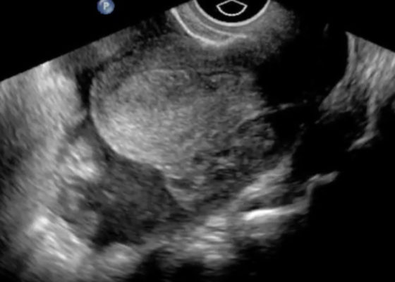
US
Ruptured Ectopic Pregnancy
DOI: https://doi.org/10.21980/J8SG6TThe patient’s serum beta-hCG was 5,637 mIU/mL. The transvaginal ultrasound showed an empty uterus with free fluid posteriorly in the pelvis and Pouch of Douglas (00:00). A 4.5 cm heterogeneous mass was visible in the left adnexa concerning for an ectopic pregnancy (00:10).
Large Right Pleural Effusion
DOI: https://doi.org/10.21980/J8D59FChest x-ray and bedside ultrasound revealed a large right pleural effusion, estimated to be greater than two and a half liters in size.
Morel-Lavallée Lesion
DOI: https://doi.org/10.21980/J88G65On physical examination, the patient was noted to have a nearly “watermelon-sized” fluctuant mass to his right lateral superior quadriceps with multiple overlying abrasions (Image 1). Computed tomography (CT) scans of the area showed a large heterogeneous collection measuring roughly 37x9.5x16 centimeters in the subcutaneous adipose layer of the lateral right thigh (Image 2), while ultrasonography revealed a complex fluid collection containing some nodular solid components and debris (Image 3). Additionally, radiographs confirmed multiple fractures including most significantly a pelvic ring fracture. Surgical debridement, evacuation, and sclerodhesis were performed nine weeks post injury to allow overlying abrasions to heal prior to intervention.
Atrial Myxoma
DOI: https://doi.org/10.21980/J87P45Bedside ultrasound revealed the presence of a left atrial mass that appeared to be tethered to the mitral valve. The mass was best viewed on ultrasound in the apical four-chamber window with the phased array probe placed over the patients’ point of maximal impact (PMI), with the patient in left lateral decubitus position.


