• Registration
  • Login
JETem
  • Home
  • About
    • Aim and Scope
    • Our Team
    • Editorial Board
    • FAQ
  • Issues
    • Current Issue
    • Ahead of Print
    • Past Issues
  • Visual EM
    • Latest Visual EM
    • Search Visual EM
    • Thumbnail Library
  • For Authors
    • Instructions for Authors
    • Submit to JETem
    • Photo Consent
    • Policies
      • Peer Review Policy
      • Copyright Policy
      • Editorial Policy, Ethics and Responsibilities
      • Conflicts of Interest & Informed Consent
      • Open Access Policy
  • For Reviewers
    • Instructions for JETem Reviewers
    • Interested in Being a JETem Reviewer?
  • Topic
    • Abdominal / Gastroenterology
    • Administration
    • Board Review
    • Cardiology / Vascular
    • Clinical Informatics, Telehealth and Technology
    • Dermatology
    • EMS
    • Endocrine
    • ENT
    • Faculty Development
    • Genitourinary
    • Geriatrics
    • Hematology / Oncology
    • Infectious Disease
    • Miscellaneous
    • Neurology
    • Ob / Gyn
    • Ophthalmology
    • Orthopedics
    • Pediatrics
    • Procedures
    • Psychiatry
    • Renal / Electrolytes
    • Respiratory
    • Toxicology
    • Trauma
    • Ultrasound
    • Wellness
    • Wilderness
  • Modality
    • Curricula
    • Innovations
    • Lectures
    • Oral Boards
    • Podcasts
    • Simulation
    • Small Group Learning
    • Team Based Learning
    • Visual EM
  • Contact Us

Large Right Pleural Effusion

Robert Rowe, MD* and Alisa Wray, MD*

*University of California, Irvine, Department of Emergency Medicine, Orange, CA

Correspondence should be addressed to Robert Rowe, MD at rower@uci.edu

DOI: https://doi.org/10.21980/J8D59FIssue 1:2
RespiratoryVisual EM
No ratings yet.
Large Right Pleural Effusion
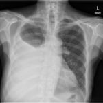
Pleural Effusion, AP Chest Xray JETem 2016


History of present illness:

An 83-year-old male with a distant history of tuberculosis status post treatment and resection approximately fifty years prior presented with two days of worsening shortness of breath. He denied any chest pain, and reported his shortness of breath was worse with exertion and lying flat.

Significant findings:

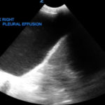
Chest x-ray and bedside ultrasound revealed a large right pleural effusion, estimated to be greater than two and a half liters in size. 

Discussion:

The incidence of pleural effusion is estimated to be at least 1.5 million cases annually in the United States.1 Erect posteroanterior and lateral chest radiography remains the mainstay for diagnosis of a pleural effusion; on upright chest radiography small effusions (>400cc) will blunt the costophrenic angles, and as the size of an effusion grows it will begin to obscure the hemidiphragm.1 Large effusions will cause mediastinal shift away from the affected side (seen in effusions >1000cc).1 Lateral decubitus chest radiography can detect effusions greater than 50cc.1

Ultrasonography can help differentiate large pulmonary masses from effusions and can be instrumental in guiding thoracentesis.1 The patient above was comfortable at rest and was admitted for a non-emergent thoracentesis. The pulmonology team removed 2500cc of fluid, and unfortunately the patient subsequently developed re-expansion pulmonary edema and pneumothorax ex-vacuo. It is generally recommended that no more than 1500cc be removed to minimize the risk of re-expansion pulmonary edema.2

Topics:

Ultrasound, POCUS, pleural effusion, chest xray, xray, lung, pulmonary, fluid overload.

References:

  1. Nicks BA, Manthey DE. Pleural effusion. In: Adams JG, Barton ED, Collings JL, DeBlieux PM, Gisondi MA, Nadel ES, eds. Emergency Medicine: Clinical Essentials. 2nd Philadelphia, PA: Elsevier; 2013:431-437.
  2. Roberts J, Hedges J. Thoracentesis. In: Adams JG, Barton ED, Collings JL, DeBlieux PM, Gisondi MA, Nadel ES, eds. Emergency Medicine: Clinical Essentials. 2nd Philadelphia, PA: Elsevier; 2013:173-189.
Icon

Pleural Effusion - Case Report

1 file(s) 637 KB
Download
Icon

Pleural Effusion - Images

1 file(s) 4.0 MB
Download
Issue 1:2USX-Ray

Reviews:

No ratings yet.

Please rate this


Morel Lavalee Lesion

Morel-Lavallée Lesion

08 Sep, 16
5th Finger Dislocation Photograph JETem 2016

Open Dislocation of Fifth Digit

08 Sep, 16
JETem is an online, open access, peer-reviewed journal-repository for EM educators

Most Viewed

  • Telemedicine Consult for Shortness of Breath Due to Sympathetic Crashing Acute Pulmonary Edema
  • Anticholinergic Toxicity in the Emergency Department
  • The Suicidal Patient in the Emergency Department Team-Based Learning Activity
  • Child Maltreatment Education: Utilizing an Escape Room Activity to Engage Learners on a Sensitive Topic
  • Acute Chest Syndrome

Visit Our Collaborators

About

Education

Learners should benefit from active learning. JETem accepts submissions of team-based learning, small group learning, simulation, podcasts, lectures, innovations, curricula, question sets, and visualEM.

Scholarship

We believe educators should advance through the scholarship of their educational work. JETem gives educators the opportunity to publish scholarly academic work so that it may be widely distributed, thereby increasing the significance of their results.

Links

  • Home
  • Aim and Scope
  • Current Issue
  • For Reviewers
  • Instructions for Authors
  • Contact Us

Newsletter

Sign up to receive updates from JETem regarding newly published issues and findings.

Copyright © 2016 JETem. All rights reserved.