• Registration
  • Login
JETem
  • Home
  • About
    • Aim and Scope
    • Our Team
    • Editorial Board
    • FAQ
  • Issues
    • Current Issue
    • Ahead of Print
    • Past Issues
  • Visual EM
    • Latest Visual EM
    • Search Visual EM
    • Thumbnail Library
  • For Authors
    • Instructions for Authors
    • Submit to JETem
    • Photo Consent
    • Policies
      • Peer Review Policy
      • Copyright Policy
      • Editorial Policy, Ethics and Responsibilities
      • Conflicts of Interest & Informed Consent
      • Open Access Policy
  • For Reviewers
    • Instructions for JETem Reviewers
    • Interested in Being a JETem Reviewer?
  • Topic
    • Abdominal / Gastroenterology
    • Administration
    • Board Review
    • Cardiology / Vascular
    • Clinical Informatics, Telehealth and Technology
    • Dermatology
    • EMS
    • Endocrine
    • ENT
    • Faculty Development
    • Genitourinary
    • Geriatrics
    • Hematology / Oncology
    • Infectious Disease
    • Miscellaneous
    • Neurology
    • Ob / Gyn
    • Ophthalmology
    • Orthopedics
    • Pediatrics
    • Pharmacology
    • Procedures
    • Psychiatry
    • Renal / Electrolytes
    • Respiratory
    • Social Determinants of Health
    • Toxicology
    • Trauma
    • Ultrasound
    • Urology
    • Wellness
    • Wilderness
  • Modality
    • Curricula
    • Innovations
    • Lectures
    • Oral Boards
      • Structured Interview
      • Communication Case
    • Podcasts
    • Simulation
    • Small Group Learning
    • Team Based Learning
    • Visual EM
  • Contact Us

Issue 2:3

Creative Commons images

Ruptured Abdominal Aortic Aneurysm

Jessica Andrusaitis, BS, MS* and Jonathan Peña, MD*

DOI: https://doi.org/10.21980/J8FP6SIssue 2:3 No ratings yet.
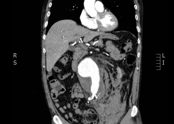
CTA demonstrated a ruptured 7.4 cm infrarenal abdominal aortic aneurysm with a large left retroperitoneal hematoma.
Cardiology/VascularVisual EM
Creative Commons images

Perforated Gastric Ulcer with Intra-abdominal Abscess

Leslie Palmerlee, MD, MPH*, Scott Mackey, DO* and Michael Petrauskis, MD, MEd*

DOI: https://doi.org/10.21980/J82H0C Issue 2:3 No ratings yet.
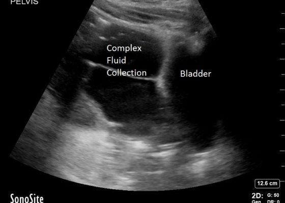
Bedside ultrasound revealed a large volume of free fluid in the right upper quadrant and in the pelvis. The fluid appeared complex with multiple septations. Its appearance was not consistent with ascites or acute intra-abdominal free fluid due to striations and pockets.
Abdominal/GastroenterologyVisual EM
Creative Commons images

Use of Bedside Compression Ultrasonography for Diagnosis of Deep Venous Thrombosis

Mohamad Moussa, MD* and Maher Abdo, BS*

DOI: https://doi.org/10.21980/J81G94 Issue 2:3 No ratings yet.
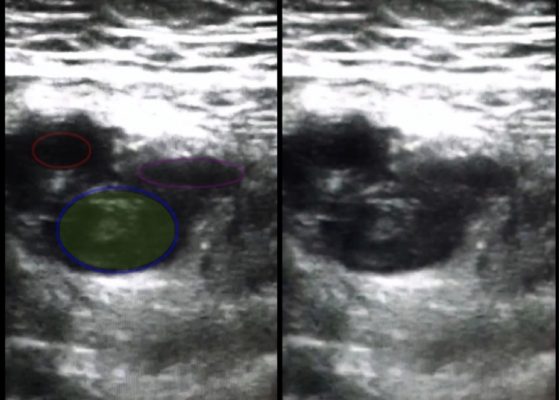
As shown in the still image of the performed ultrasound, a transverse view of the proximal-thigh revealed a visible thrombus (green shading) occluding the lumen of the left common femoral vein (blue ring), which was non-compressible when direct pressure was applied to the probe. Also visible is a patent and compressible branch of the common femoral vein (purple ring) and the femoral artery (red ring), highlighted by its thick vessel wall and pulsatile motion.
UltrasoundCardiology/VascularVisual EM
Creative Commons images

Open Pneumothorax

Bart Paull, MD*, Zachary Jarou, MD* and David Richards, MD*

DOI: https://doi.org/10.21980/J88036 Issue 2:3 No ratings yet.
A large chest wound was clinically obvious. A chest radiograph performed after intubation showed subcutaneous emphysema, an anterior rib fracture, and a right-sided pneumothorax. He was then taken to the operating room for further management.
TraumaRespiratoryVisual EM
Creative Commons images

Dense MCA Sign

Richard J Chen, MD* and Grant Wei, MD*

DOI: https://doi.org/10.21980/J8CS66Issue 2:3 No ratings yet.
A non-contrast computed tomography (CT) scan showed a hyperdensity along the right middle cerebral artery (MCA) consistent with acute thrombus. The red arrow highlights the hyperdensity in the annotated image.
NeurologyVisual EM
Creative Commons images

Supracondylar Fracture

Jessica Andrusaitis, BS, MS* and Ben Feldman, MD*

DOI: https://doi.org/10.21980/J8492P Issue 2:3 No ratings yet.
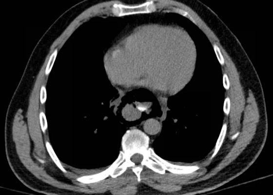
History of present illness: A 15-year-old male presented to the emergency department with right elbow pain after falling off a skateboard.  The patient denied a decrease in strength or sensation but did endorse paresthesias to his hand.  On exam, the patient had an obvious deformity of his right elbow with tenderness to palpation and decreased range of motion at the
OrthopedicsVisual EM
«‹3
Page 3 of 3

JETem is an online, open access, peer-reviewed, journal-repository for EM educators. We are PMC Indexed.

Most Viewed

  • The Silent Saboteur: Teaching the Clinical Implications of Occult Hypoxemia & Social Determinants of Health via a Pulmonary Embolism Case
  • Diabetic Ketoacidosis and Necrotizing Soft Tissue Infection
  • My Broken Heart
  • Stabilization of Cardiogenic Shock for Critical Care Transport, a Simulation
  • Innovative Ultrasound-Guided Erector Spinae Plane Nerve Block Model for Training Emergency Medicine Physicians

Visit Our Collaborators

Creative Commons Licence
This work is licensed under a Creative Commons Attribution 4.0 International License.

About

Education

Learners should benefit from active learning. JETem accepts submissions of team-based learning, small group learning, simulation, podcasts, lectures, innovations, curricula, question sets, and visualEM.

Scholarship

We believe educators should advance through the scholarship of their educational work. JETem gives educators the opportunity to publish scholarly academic work so that it may be widely distributed, thereby increasing the significance of their results.

Links

  • Home
  • Aim and Scope
  • Current Issue
  • For Reviewers
  • Instructions for Authors
  • Contact Us

Newsletter

Sign up to receive updates from JETem regarding newly published issues and findings.

Copyright Creative Commons Attribution 4.0 International