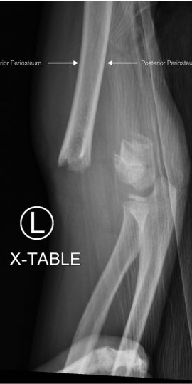
Pediatric Supracondylar Fracture
History of present illness:
A 7-year-old left-handed male presented with left arm pain and deformity after being tackled while playing. On exam, there appeared to be dorsal displacement of the distal segment of the upper extremity. He had two-plus radial and ulnar pulses, and normal capillary refill. Sensation was intact to axillary, radial, ulnar, and median nerve distributions. Compartments were soft.
Significant findings:
Plain film radiography showed a displaced supracondylar fracture with disrupted anterior and posterior periostea, consistent with a type 3 supracondylar fracture.
Discussion:
Supracondylar fractures are the most common pediatric elbow fracture.1 Approximately 95% are due to a fall onto an outstretched hand while the elbow is in extension. Direct trauma to the posterior aspect of a flexed elbow accounts for the remainder.2 There are three classifications of supracondylar fractures: type 1 is non-displaced, type 2 is displaced, but has an intact posterior periosteum, and type 3 is displaced with disrupted anterior and posterior periostea.
Careful examination assessing for pulses, perfusion, neurologic integrity, and elevated compartment pressures are important in the evaluation.3 The brachial artery is often injured in posterior lateral displaced fractures.4 Neurologic deficits to the median, ulnar, or radial nerves are seen in as many of 49% of Type 3 supracondylar fractures; however, neuropraxias often resolve within two to three months.5 Untimely treated compartment syndrome may lead to Volkmann ischemic contractors, which are characterized by flexion of the elbow, pronation of the forearm, flexion of the wrist, and extension of the metacarpal phalangeal joints.6
Plain film radiography oriented in the anterior-posterior (AP) and lateral fashions are typically sufficient for diagnosis; however, a fracture may exist without overt signs on X-ray.7
Given the high morbidity associated with Type 3 fractures, emergent Orthopedic consult is required because surgical intervention is required.1
Topics:
Orthopedics, ortho, elbow, supracondylar facture, peds, pediatrics.
References:
- Carson S, Woolridge DP, Colletti J, Kilgore K. Pediatric upper extremity injuries. Pediatr Clin North Am. 2006;53(1):41-67. doi: 10.1016/j.pcl.2005.10.003
- Baratz M, Micucci C, Sangimino M. Pediatric supracondylar humerus fractures. Hand Clin. 2006;22(1):69-75. doi: 10.1016/j.hcl.2005.11.002
- Jones ET, Louis DS. Median nerve injuries associated with supracondylar fractures of the humerus in children. Clin Orthop Relat 1980;(150):181-186.
- Griffin KJ, Walsh SR, Markar S, Tang TY, Boyle JR, Hayes PD. The pink pulseless hand: a review of the literature regarding management of vascular complications of supracondylar humeral fractures in children.Eur J Vasc Endovasc Surg. 2008;36:697-702. doi: 10.1016/j.jvs.2008.10.054
- Campbell CC, Waters PM, Emans JB, Kasser JR, Millis MB. Neurovascular injury and displacement in type III supracondylar humerus fractures. J Pediatr Orthop. 1995;15(1):47-52.
- Wu J, Perron AD, Miller MD, Powell SM, Brady WJ. Orthopedic pitfalls in the ED: pediatric supracondylar humerus fractures. Am J Emerg Med. 2002;20(6):544-550.
- Rajiah P. Surgical Radiology: Clinical Cases. Cheshire, UK: PasTest Ltd. 2007;317.


