Right Atrial Thrombus
History of present illness:
A 77-year-old male presented to the emergency department with shortness of breath. Symptoms progressively worsened over the last 4-5 days, and on arrival was associated with chest tightness. He denied any medical conditions, smoking, or pertinent family history. He has not seen a primary care physician in “many years.” Upon arrival he was in mild distress. On exam, patient was tachypneic, tachycardic, and hypoxic with clear lung sounds. His electrocardiogram demonstrated sinus tachycardia.
Significant findings:
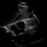
Bedside echocardiogram was performed, which revealed a free-floating thrombus in the right atrium on the sub-xiphoid view as seen in the video. The right atrium is denoted by the blue circle, in which a hyperechoic mobile mass can be seen. This finding was confirmed by an official echocardiogram which shows the thrombus in the right atrium extending through the tricuspid valve, as shown in the second image denoted by the red arrow. Significant right heart strain was also found, with severe pulmonary hypertension and intraventricular septal flattening.
Discussion:
Right atrial thrombus is a rare finding that is almost exclusively found complicating pulmonary thromboembolism1 and carries a high mortality rate from 27%1 to 44%.2 Typically the diagnosis is made via echocardiogram, with the sub-xiphoid view most likely to provide adequate views of the right atrium. The sub-xiphoid view is obtained by using the left lobe of the liver as an acoustic window, with the probe angled cephalad toward the thorax. Treatment strategies for right atrial thrombus include anticoagulation, surgical thrombectomy, or thrombolytic therapy. At this time there are no evidenced-based guidelines to direct treatment strategy; however, the largest retrospective analysis shows significantly increased survival with thrombolysis, having a mortality rate of 11.3%, in comparison to 28.6% with anticoagulation and 23.8% with surgical thrombectomy.3 Another study reports that hypotension and pulmonary embolism severity predicts mortality risk in the setting of right heart thrombi, but not the size, shape, or mobility of the thrombus on echocardiogram.4
Our patient was immediately started on anticoagulation, and a computed tomography (CT) scan was also performed and showed significant bilateral pulmonary emboli. Several hours after arrival he developed worsening dyspnea and hypoxia. He was treated with an intravenous bolus of tissue plasminogen activator (tPA). Shortly afterwards he had two episodes of cardiac arrest with pulseless electrical activity, with return of spontaneous circulation after epinephrine and sodium bicarbonate administration. His clinical course was further complicated by anemia secondary to gastrointestinal bleeding, and at that time a Greenfield filter was placed while anticoagulation was held. He was neurologically intact and discharged home eight days after presentation on coumadin for anticoagulation.
Topics:
Right atrial thrombus, cardiac thrombus, echocardiogram, pulmonary embolism.
References:
- Torbiki A, Galie N, Covezzoli A, et al. Right heart thrombi in pulmonary embolism: results from the international cooperative pulmonary embolism registry. J Am Coll Cardiol. 2003;41(12):2245-2251.
- Chartier L, Béra J, Delomez, M, et al. Free-floating thrombi in the right heart: diagnosis, management, and prognostic indexes in 38 consecutive patients. Circulation. 1999;99(21):2779-2783.
- Rose PS, Punjabi NM, Pearse DB. Treatment of right heart thromboemboli. Chest. 2002; 121(13):806-814.
- Elikowski W, Grifoni S, Hugues T. Outcome of patients with right heart thrombi: the right Heart Thrombi European Registry. Eur Respir J. 2016;47(3):869-875. doi: 10.1183/13993003.00819-2015.


