Perforated Duodenal Ulcer
History of present illness:
A 53-year-old male with a history of daily alcohol abuse presented with sudden onset epigastric pain. The pain radiated to the right upper abdominal quadrant and was associated with shortness of breath and nausea. The patient’s vitals were notable for blood pressure of 181/107 and a heart rate of 124. He was in moderate distress and had a firm, distended abdomen with diffuse tenderness to palpation, without rebound or guarding.
Significant findings:
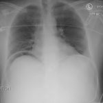
In the chest radiograph, there was obvious free air under the both the right diaphragm (above the liver) and the left diaphragm, consistent with pneumoperitoneum.
Discussion:
A perforated ulcer is a surgical emergency. Overall mortality has been shown to be approximately 6.2%.1 Rapid diagnosis is essential as prognosis improves if treatment is initiated within the first six hours and worsens after 12 hours.2 The sensitivity for detecting pneumoperitoneum on plain radiography ranges from 50%-80%3-8 with specificity of 53%.7 An upright chest radiograph can detect as little as one to two milliliters of air.9,10 If free air is not seen on a posteroanterior (PA) upright chest radiograph, an upright lateral chest radiograph can be obtained, which is more sensitive (98% sensitivity).8,11 About 10%-20% of ruptured ulcers will not present with visible free-air under the diaphragm on plain x-ray.12 In this case, given the free air seen on chest radiograph and peritoneal signs on exam, the patient was taken straight to the operating room for general surgery.
Topics:
Perforated ulcer, pneumoperitoneum, gastroenterology, GI, x-ray, radiograph.
References:
- Sung JJ, Lau JY, Ching JY, Wu JC, Lee YT, Chiu PW, et al. Continuation of low-dose aspirin therapy in peptic ulcer bleeding: a randomized trial. Ann Intern Med. 2010;152(1):1-9. doi: 10.7326/0003-4819-152-1-201001050-00179
- Svanes C, Lie RT, Svanes K, Lie SA, Søreide O. Adverse effects of delayed treatment for perforated peptic ulcer. Ann Surg. 1994;220(2):168-175.
- Furukawa A, Sakoda M, Yamasaki M, et al. Gastrointestinal tract perforation: CT diagnosis of presence, site, and cause. Abdom Imaging. 2005;30(5):524-534. doi: 10.1007/s00261-004-0289-x
- Cho KC, Baker SR. Extraluminal air. Diagnosis and significance. Radiol Clin North Am. 1994;32(5):829-844.
- Ghahremani GG. Radiologic evaluation of suspected gastrointestinal perforations. Radiol Clin North Am. 1993;31(6):1219-1234.
- Maniatis V, Chryssikopoulos H, Roussakis A, Kalamara C, Kavadias S, Papadopoulos A, et al. Perforation of the alimentary tract: evaluation with computed tomography. Abdom Imaging. 2000;25(4):373-379.
- Gans SL, StokerJ, Boermeester, MA. Plain abdominal radiography in acute abdominal pain; past, present, and future. Int J Gen Med. 2012;5:525-533. doi: 10.2147/IJGM.S17410
- Woodring JH, Heiser MJ. Detection of pneumoperitoneum on chest radiographs: comparison of upright lateral and posteroanterior projections. AJR Am J Roentgenol. 1995;165(1):45-47. doi: 10.2214/ajr.165.1.7785629
- Billittier AJ, Abrams BJ, Brunetto A. Radiographic imaging modalities for the patient in the emergency department with abdominal complaints. Emerg Med Clin North Am. 1996;14(4):789-850. doi: 1016/S0733-8627(05)70279-0
- Mindelzun RE, Jeffrey RB. Unenhanced helical CT for evaluating acute abdominal pain: a little more cost, a lot more information. Radiology. 1997;205(1):43-45. doi: 10.1148/radiology.205.1.9314959
- Markowitz SK, Ziter FM Jr. The lateral chest film and pneumoperitoneum. Ann Emerg Med. 1986;15(4):425-427.
- Grassi R, Romano S, Pinto A, Romano L. Gastro-duodenal perforations: conventional plain film, US and CT findings in 166 consecutive patients. Eur J Radiol. 2004;50(1):30-36. doi: 10.1016/j.ejrad.2003.11.012


