Gastric Volvulus
History of present illness:
An 83-year-old male without significant medical history presented to the emergency department with the complaint of 40 pound weight loss in the last four months and significant abdominal pain and distention starting two days prior to presentation. The patient had been having decreased appetite without any episodes of vomiting, diarrhea, blood in stools.
In the emergency department, the patient appeared cachectic and ill. Physical exam showed distended abdomen and significant pain in the epigastric region with associated guarding with deep palpation of his abdominal region.
Significant findings:
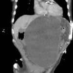
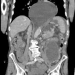
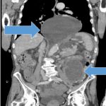
Point of care ultrasound of his abdomen showed a large fluid filled structure with well-defined borders containing gastric contents extending from the xiphoid process to the umbilical region. No free fluid was noted on focus assessment with sonography for trauma (FAST) examination. A computed tomography (CT) scan was performed emergently and it was noted that the patient had a significantly distended stomach and gastric volvulus (blue arrows) noted in the area of his paraesophageal/hiatal hernia.
Discussion:
Gastric Volvulus is caused by an abnormal rotation of the stomach more than 180 degrees, creating a closed loop obstruction. Borchardt’s triad, which is severe epigastric pain, retching without vomiting and inability to pass nasogastric tube, has been seen in 70 percent of cases.1
The most common cause of gastric volvulus in adults is from diaphragmatic defects caused by paraesophageal or hiatal hernia. Gastric volvulus is the most common complication of paraesophageal hernias.2 Males and females are equally affected, while 10-20 percent of cases occur in children, most commonly less than one year old and has higher incidence in patients older than 50 years old.1 Based on the age of the patient plus the CT scan findings, the patient had a type 2 acquired gastric volvulus, secondary to a hiatal vs paraesophageal hernia, which is the most common type occurring in 59% of cases.2 A type 1 gastric volvulus is idiopathic due to abnormal laxity of the peritoneal ligaments of the stomach. A type 2 gastric volvulus is a congenital or acquired anatomical defect that allows the stomach increased mobility.3
Upper gastrointestinal barium studies can be used to diagnose gastric volvulus with a diagnostic yield of 81%-84%.4 Ultrasound can be used to help diagnose gastric volvulus but there have been no large studies performed to determine the sensitivity and specificity of ultrasound, with conflicting case reports showing normal ultrasound findings vs a single case report providing evidence of gastric volvulus. Computed tomography can be used and the presence of two findings on CT scan makes the diagnosis of gastric volvulus 100% sensitive and specific. These findings are: the antropyloric transition point is located without any abnormalities in the transition zone and the antrum is at the same level or higher than the fundus.5
Surgery is the definitive management for gastric volvulus with nasogastric tube placement if possible.The prognosis for non-operative patients is poor with mortality as high as 80 percent due to the volvulus causing strangulation, necrosis and perforation. With surgical management the mortality is 15-20 percent.6
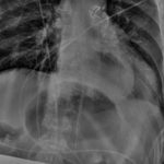
In this case, the patient elected for his code status to be do not resuscitate – comfort care. The patient had a nasogastric tube placed with the return of 3000ml of dark fluid within minutes of placement, which resulted in near complete relief of his abdominal pain and distention (blue arrows on chest x-ray). He was admitted to the hospice service and died three days later.
Key points that should be applied to clinical practice are that a gastric volvulus should be promptly recognized and diagnosed since any delay could cause increased mortality in a surgical emergency that already carries a high mortality, and that point of care ultrasound is a valuable tool to help guide management and treatment of abdominal pathology. This case report raises the question of how well ultrasound can help with the diagnosis of gastric volvulus and the need for larger studies to determine its diagnostic value.
Topics:
Gastric Volvulus, Borchardt’s triad, abdominal pain.
References:
- Rashid, F, Thangarajah, T, Mulvey, D, Larvin, M, Iftikhar SY. A review article on gastric volvulus: a challenge to diagnosis and management. Int J Surg. 2010;8(1):18-24. doi: 10.1016/j.ijsu.2009.11.002.
- Shivanand G, Seema S, Srivastava DN, et al. Gastric volvulus: acute and chronic presentation. Clin Imaging. 2003 27(4):265-268.
- Miller DL, Pasquale MD, Seneca RP, Hodin E. Gastric volvulus in the pediatric population. Arch Surg. 1991: 126(9):1146-1149.
- Gourgiotis S, Vougas V, Germanos S, Baratsis, S. Acute gastric volvulus: diagnosis and management over 10 years. Dig Surg. 2006: 23(3):169-172.
- Millet I, Orliac C, Alili C, Guillon F, Taourel P. Computed tomography findings of acute gastric volvulus. Eur Radiol. 2014: (12):3115-3122. doi: 10.1007/s00330-014-3319-2.
- Palanivelu C, Rangarajan M, Shetty AR, Senthilkumar R. Laparoscopic suture gastropexy for gastric volvulus: a report of 14 cases. Surg Endosc. 2007 (6):863-866.


