Ectopic Kidney
History of present illness:
A 50-year-old male with no past medical history presented to the emergency department with a chief complaint of right flank pain after stretching. His vital signs were within normal limits and physical exam was significant for tenderness to palpation over the right lateral chest wall. Chest X-ray was unremarkable. Due to the patient’s uncertainty of the exact mechanism of injury, additional trauma could not be ruled out and a bedside focused assessment with sonography in trauma (FAST) scan was performed, which was negative for free fluid, but notable for an absence of a right kidney. The patient was sent for a computed tomography (CT) abdomen/pelvis to evaluate the etiology of symptoms and to address the absence of visualized kidney on ultrasound.
Significant findings:
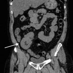
CT of the abdomen and pelvis revealed a normal left kidney and an ectopic, malrotated right kidney located in the pelvis (see white arrow).
Discussion:
Renal ectopia is described as a malposition of the kidney, due to faulty migration from the fetal pelvis during early embryonic development. Evidence suggests an incidence ranging from 1:900 to 1:12,000.1-3 While most cases are asymptomatic and do not require intervention, complications include vesicoureteral reflux, urinary tract infections, hydronephrosis, and renal calculi.4,5 Ultrasonography is indicated for the evaluation of free fluid in the abdomen and pelvis in the setting of trauma. In this case, the right upper quadrant ultrasound was negative for both free fluid and a right kidney, even with appropriate repositioning techniques.7 The absence of the right kidney on ultrasound in the setting of pain prompted the decision for further diagnostic imaging, which revealed an ectopic right kidney. The absence of a kidney on FAST exam should prompt the clinician to consider surgical (eg, nephrectomy) or congenital (eg, renal ectopy) explanations. Furthermore, pathophysiologic processes (eg, pyelonephritis) that may occur in an ectopic right kidney can masquerade as disease entities which cause right lower quadrant pain such as acute appendicitis.8
Topics:
Renal ectopy, FAST scan, trauma, CT scan.
References:
- Meizner I, Yitzhak M, Levi A, Barki Y, Barnhard Y, Glezerman, M. Fetal pelvic kidney: a challenge in prenatal diagnosis? Ultrasound Obstet Gynecol. 1995;5(6):391-3. doi: 10.1046/j.1469-0705.1995.05060391.x
- Yuksel A, Batukan C. Sonographic findings of fetuses with an empty renal fossa and normal amniotic fluid volume. Fetal Diagn Ther. 2004;19(6):525. doi: 10.1159/000080166
- Sheih C, Liu M, Hung C, Yang K, Chen W, Lin C. Renal abnormalities in schoolchildren. Pediatrics. 1989;84(6):1086-90.
- Guarino N, Tadini B, Camardi P, Silvestro L, Lace R, Bianchi M. The incidence of associated urological abnormalities in children with renal ectopia. J Urol. 2004;172(4 Pt 2):1757-9;discussion 1759. doi: 10.1097/01.ju.0000138376.93343.74
- Bhoil R, Sood D, Singh Y, Nimkar K, Shukla A. An ectopic pelvic kidney. Pol J Radiol. 2015;80:425–427. doi: 10.12659/PJR.894603
- American College of Emergency Physicians. Emergency ultrasound imaging criteria compendium. American College of Emergency Physicians. Ann Emerg Med. 2006;48(4):487-510. doi: 10.1016/j.annemergmed.2006.07.946
- Fanzen, D, Patwari R. FAST examination. Clerkship directors in emergency medicine (CDEM) curriculum website. https://cdemcurriculum.com/fast-examination/. Updated 2016. Accessed July 10, 2017.
- Lossius M, Araya C, Henry D, Neiberger R. A patient with an unusual cause right lower quadrant pain and vomiting: pyelonephritis of an ectopic right kidney masquerading as acute appendicitis. Case Rep Med. 2009:638501. doi: 10.1155/2009/638501


