Procedural Sedation for the removal of a rectal foreign body
History of present illness:
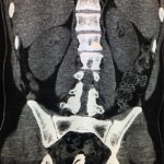
A 40-year-old male with a history of intravenous drug use presented to the emergency department (ED) for one week of constant lower abdominal pain associated with bloody stool. He denied fever, nausea, vomiting, urinary symptoms, and testicular pain or swelling. On exam, vital signs were within normal limits. Abdominal exam was non-tender without rebound or guarding. Rectal exam was negative for occult blood but positive for a palpable firm, blunt object. A computed tomography (CT) of the abdomen and pelvis was ordered to further investigate.
Significant findings:
Axial and coronal views on CT showed evidence of a large, tube-shaped foreign body in the rectum (see arrows) without evidence of acute gastrointestinal tract disease.
Discussion:
While rectal foreign bodies (RFB) are not uncommon to the ED, accurate epidemiological estimates are not available, due in part to underreporting.1 One study estimated an incidence of one patient per month that needed care for a RFB.2 Generally, patients can remove the object themselves; however, 20% of cases require endoscopic intervention and 1% require surgical intervention.3
RFBs can be removed via the transanal approach manually, instrument-assisted (eg, Kocher clamp, obstetric forceps), or endoscopically. In cases without intestinal perforation, transanal removal of a RFB is generally attempted as a first-line procedure in the ED, with an approximate success rate of 75%.4 However, the limitations of transanal removal depends on the location of the object, level of anal relaxation, and ability to grasp the object, which may be limited by the provider’s hand size and availability of instruments.Ways of facilitating bedside removal of RFBs include the Valsalva maneuver with proper positioning (i.e., lithotomy or prone knee-to-chest position) or manual abdominal wall compression to help move the object closer to the anal orifice. Anoscopy may also be used to improve visualization of the RFB. In some cases, RFBs can create a vacuum effect for which Foley catheters may be used to break the seal and provide additional traction. After the lubricated Foley passes proximal to the RFB and the balloon is inflated, gentle traction should be employed to move the object closer. Sedation can also be employed to decrease rectal tone and make the procedure more tolerable for the patient.
Complications prior to or during removal of the RFB include tearing of the rectal mucosa, perforation, infection, fecal incontinence, bladder and vessel injury or migration of the RFB to the chest wall.3,5 Uncontrollable rectal bleeding, peritonitis, or perforation are contraindications to ED RFB removal and warrant surgery or gastroenterology consultation. General anesthesia can be used for laparotomy with single incision to remove RFBs in the operating room.2
In this case, procedural sedation was utilized to facilitate removal of the object. Ketamine (2 mg/kg intravenously) was initially proposed, but due to concerns of the patient’s history of drug abuse, alternatives were considered. While Ketamine is generally well-tolerated, the incidence of dysphoric reactions has been estimated to occur in 10%-20% of patients.6 There is also some evidence that patients with a history of drug abuse may be more likely to have tolerance to Ketamine that requires higher dosing.7 Taking these factors into account, Etomidate (0.2 mg/kg intravenously) was used instead with favorable results. A metallic flashlight was removed by grasping the object with a combination of forceps and manual manipulation. After RFB removal, sigmoidoscopy was recommended to assess for rectal mucosal injuries or tears.8 No additional injuries were found on sigmoidoscopy and the patient tolerated the procedure well without complications.
Topics:
Abdominal pain, computed tomography, CT, foreign body, procedural sedation.
References:
- Tupe CL, Pham TV. Anorectal complaints in the emergency department. Emerg Med Clin North Am. 2016;34(2):251-270.doi: 1016/j.emc.2015.12.013
- Lake JP, Essani R, Petrone P, Kaiser AM, Asensio J, Beart RW. Management of retained colorectal foreign bodies: predictors of operative intervention. Dis Colon Rectum. 2004;47(10):1694-1698.
- Anderson KL, Dean AJ. Foreign bodies in the gastrointestinal tract and anorectal emergencies. Emerg Med Clin North Am. 2011;29(2):369–400. doi: 10.1016/j.emc.2011.01.009
- Mikami H, Ishimura N, Oka A, et al. Successful transanal removal of a rectal foreign body by abdominal compression under endoscopic and x-ray fluoroscopic observation: a case report. Case Rep Gastroenterol. 2016;10(3):646-652. doi:10.1159/000452210
- Goldberg JE, Steele SR. Rectal foreign bodies. Surg Clin North Am. 2010;90(1):173-184. doi: 10.1016/j.suc.2009.10.004
- Strayer RJ, Nelson LS. Adverse events associated with ketamine for procedural sedation in adults. Am J Emerg Med. 2008;26(9):985-1028. doi: 1016/j.ajem.2007.12.005
- Farkas, J. The ketamine-tolerant patient. Available on: https://emcrit.org/pulmcrit/ketamine-tolerance/. Published September 11, 2017. Accessed October 10, 2017.
- Cohen JS, Sackier JM. Management of colorectal foreign bodies. J R Coll Surg Edinb. 1996;41(5):312-315.


