Volvulus
History of present illness:
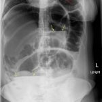
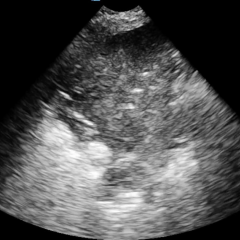
A 26-year-old previously healthy female presented to the emergency department (ED) with diffuse abdominal pain, distention, and constipation for two days. Her physical exam was normal except for a soft, distended abdomen with mild diffuse tenderness to palpation. Vital signs, labs and right upper quadrant ultrasound performed in the ED were all within normal limits. Acute abdominal series (AAS) x-ray was significant for a sigmoid volvulus. Follow up CT confirmed the findings and the patient was subsequently admitted for emergent flexible sigmoidoscopy.
Significant findings:
Upright and supine frontal radiographs of the abdomen demonstrate gas dilation of the large bowel from the level of the cecum to the sigmoid colon with air fluid levels (yellow arrows). There is a swirled configuration of the distal descending to sigmoid colon indicating the level of the volvulus (dashed yellow line) and giving rise to the classic “coffee bean” sign (dotted white tracing). Note the elevated left hemidiaphragm on the upright view reflecting abdominal distention with increased intra-abdominal pressure (red arrow).
Discussion:
Volvulus is an emergent condition that occurs when the colon twists on its mesenteric axis greater than 180 degrees, producing obstruction of intestinal lumen and mesenteric vessels.1 The incidence of volvulus is rare in the United States, with the most common locations of volvulus being the sigmoid colon and cecum with 60%-70% and 20%-30% of cases reported, respectively.2 The small remaining fraction of cases occur in the splenic flexure and transverse colon.2 Sigmoid volvulus occurs more often in elderly patients with multiple comorbidities or those with neurological and psychiatric diseases and has a more subtle and insidious clinical presentation.3 In contrast, cecal volvulus is characterized by acute onset and is most frequently seen in younger (25-35), healthier individuals, particularly in long distance runners or patients who have had previous abdominal surgeries.4
Abdominal radiographs may assist with diagnosis; however CT, magnetic resonance imaging, and flexible imaging are more accurate.5 Flexible sigmoidoscopy is the standard procedure for patients with viable bowel, and sudden decompression at rigid sigmoidoscopy is successful in 70-90% of cases.2 However, for patients who fail decompression or present with diffuse peritonitis, intestinal perforation, or ischemic necrosis, emergency surgery is the appropriate treatment.2 Timely identification and management are crucial in treating sigmoid volvulus before the appearance of these aforementioned life-threatening complications.6
Topics:
volvulus, sigmoidoscopy, gastroenterology, constipation.
References:
- Gerwig WH. Volvulus of the colon: symposium on function and disease of anorectum and colon.Surg Clin North Am. 1950;60(4):721-742. doi: 1001/archsurg.1950.01250010742008
- Lou Z, Yu ED, Zhang W, Meng RG, Hao LQ, Fu GC. Appropriate treatment of acute sigmoid volvulus in the emergency setting. World J Gastroenterol.2013;19(30):4979–4983. doi: 10.3748/wjg.v19.i30.4979
- Madiba TE, Thomson SR. The management of sigmoid volvulus. J R Coll Surg Edinb. 2000;45:74-80.
- Mulas C, Bruna M, García-Armengol J, Roig JV. Management of colonic volvulus. Experience in 75 patients. Rev Esp Enferm Dig.2010;102:239-248. doi: 10.4321/S1130-01082010000400004
- Young WS, Engelbrecht HE, Stocker A. Plain film analysis in sigmoid volvulus. Clin Radiol.1978;29:553-560. doi: 10.1016/S0009-9260(78)80049-X
- Katsikogiannis N, Machairiotis N, Zarogoulidis P, Sarika E, Stylianaki A, Zisoglou M, et al Management of sigmoid volvulus avoiding sigmoid resection. Case Rep Gastroenterol.2012;6:293-299. doi: 10.1159/000339216


