• Registration
  • Login
JETem
  • Home
  • About
    • Aim and Scope
    • Our Team
    • Editorial Board
    • FAQ
  • Issues
    • Current Issue
    • Ahead of Print
    • Past Issues
  • Visual EM
    • Latest Visual EM
    • Search Visual EM
    • Thumbnail Library
  • For Authors
    • Instructions for Authors
    • Submit to JETem
    • Photo Consent
    • Policies
      • Peer Review Policy
      • Copyright Policy
      • Editorial Policy, Ethics and Responsibilities
      • Conflicts of Interest & Informed Consent
      • Open Access Policy
  • For Reviewers
    • Instructions for JETem Reviewers
    • Interested in Being a JETem Reviewer?
  • Topic
    • Abdominal / Gastroenterology
    • Administration
    • Board Review
    • Cardiology / Vascular
    • Clinical Informatics, Telehealth and Technology
    • Dermatology
    • EMS
    • Endocrine
    • ENT
    • Faculty Development
    • Genitourinary
    • Geriatrics
    • Hematology / Oncology
    • Infectious Disease
    • Miscellaneous
    • Neurology
    • Ob / Gyn
    • Ophthalmology
    • Orthopedics
    • Pediatrics
    • Pharmacology
    • Procedures
    • Psychiatry
    • Renal / Electrolytes
    • Respiratory
    • Social Determinants of Health
    • Toxicology
    • Trauma
    • Ultrasound
    • Urology
    • Wellness
    • Wilderness
  • Modality
    • Curricula
    • Innovations
    • Lectures
    • Oral Boards
      • Structured Interview
      • Communication Case
    • Podcasts
    • Simulation
    • Small Group Learning
    • Team Based Learning
    • Visual EM
  • Contact Us

Globe Rupture

Reid Honda, MD*

*University of California, Irvine, Department of Emergency Medicine, Orange, CA

Correspondence should be addressed to Reid Honda, MD at rkhonda@uci.edu  

DOI: https://doi.org/10.21980/J8N91Z Issue 2:3
OphthalmologyVisual EM
No ratings yet.
Creative Commons images

History of present illness:

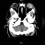
A 46-year-old male presented to the emergency department (ED) with severe left eye pain and decreased vision after tripping and striking the left side of his head on the corner of his wooden nightstand.  The patient arrived as an inter-facility transfer for a suspected globe rupture with a protective eye covering in place; thus, further physical examination of the eye was not performed by the emergency physician in order to avoid further leakage of aqueous humor.

Significant findings:

The patient’s computed tomography (CT) head demonstrated a deformed left globe, concerning for ruptured globe. The patient had hyperdense material in the posterior segment (see green arrow), consistent with vitreous hemorrhage. CT findings that are consistent with globe rupture may include a collapsed globe, intraocular air, or foreign bodies.

Discussion:

A globe rupture is a full-thickness defect in the cornea, sclera, or both.1 It is an ophthalmologic emergency. Globe ruptures are almost always secondary to direct perforation via a penetrating mechanism; however, it can occur due to blunt injury if the force generated creates sufficient intraocular pressure to tear the sclera.2 Globes most commonly rupture at the insertions of the intraocular muscles or at the limbus. They are associated with a high rate of concomitant orbital floor fractures.2,3 Possible physical examination findings include a shallow anterior chamber on slit-lamp exam, hyphema, and an irregular “teardrop” pupil. Additionally, a positive Seidel sign, which is performed by instilling fluorescein in the eye and then examining for a dark stream of aqueous humor, is indicative of a globe rupture.4 CT is often used to assess for globe rupture; finds of a foreign body, intraocular air, abnormal contour or volume of the globe, or disruption of the sclera suggest globe rupture.2 The sensitivity of CT scan for diagnosis of globe rupture is only 75%; thus, high clinical suspicion is needed. CT should only be used as an adjunct for physical exam.3

Direct pressure (including tonometry measurements) should never be applied to a globe that is suspected or confirmed to be ruptured because of the risk for extrusion of the intraocular contents.3 Antibiotics should be given to prevent endopthalmitis. Antiemetics should also be given to prevent Valsalva maneuvers which can worsen the globe rupture.3 Ophthalmologic consultation for surgery should be obtained emergently.5

Topics:

Ophthalmology, globe rupture, globe perforation.

References:

  1. Bhatia K, Sharma R. Eye emergencies. In: Adams JG, Barton ED, Collings JL, DeBlieux PMC, Gisondi MA, Nadel ES, eds. Emergency Medicine Clinical Essentials. 2nd Philadelphia, PA: Elsevier Saunders; 2013:224.
  2. Dunkin JM, Crum AV, Swanger RS, Bokhari SAJ. Globe trauma. YSULT. 2011;32(1):51-56. doi: 10.1053/j.sult.2010.09.003
  3. Bord SP, Linden J. Trauma to the globe and orbit.Emerg Med Clin North Am. 2008;26(1):97-123. doi:10.1016/j.emc.2007.11.006
  4. Babineau MR, Sanchez LD. Ophthalmologic procedures in the emergency department. Emerg Med Clin North. 2008;26(1):17-34. doi: 10.1016/j.emc.2007.11.003
Icon

Globe Rupture - Case Report

1 file(s) 298 KB
Download
Icon

Globe Rupture - Images

1 file(s) 157 KB
Download
CTIssue 2:3

Reviews:

No ratings yet.

Please rate this


Creative Commons images

Steven-Johnson Syndrome

04 Jul, 17
Creative Commons images

Esophageal Perforation

04 Jul, 17

JETem is an online, open access, peer-reviewed, journal-repository for EM educators. We are PMC Indexed.

Most Viewed

  • The Silent Saboteur: Teaching the Clinical Implications of Occult Hypoxemia & Social Determinants of Health via a Pulmonary Embolism Case
  • Diabetic Ketoacidosis and Necrotizing Soft Tissue Infection
  • My Broken Heart
  • Stabilization of Cardiogenic Shock for Critical Care Transport, a Simulation
  • Innovative Ultrasound-Guided Erector Spinae Plane Nerve Block Model for Training Emergency Medicine Physicians

Visit Our Collaborators

Creative Commons Licence
This work is licensed under a Creative Commons Attribution 4.0 International License.

About

Education

Learners should benefit from active learning. JETem accepts submissions of team-based learning, small group learning, simulation, podcasts, lectures, innovations, curricula, question sets, and visualEM.

Scholarship

We believe educators should advance through the scholarship of their educational work. JETem gives educators the opportunity to publish scholarly academic work so that it may be widely distributed, thereby increasing the significance of their results.

Links

  • Home
  • Aim and Scope
  • Current Issue
  • For Reviewers
  • Instructions for Authors
  • Contact Us

Newsletter

Sign up to receive updates from JETem regarding newly published issues and findings.

Copyright Creative Commons Attribution 4.0 International