• Registration
  • Login
JETem
  • Home
  • About
    • Aim and Scope
    • Our Team
    • Editorial Board
    • FAQ
  • Issues
    • Current Issue
    • Ahead of Print
    • Past Issues
  • Visual EM
    • Latest Visual EM
    • Search Visual EM
    • Thumbnail Library
  • For Authors
    • Instructions for Authors
    • Submit to JETem
    • Photo Consent
    • Policies
      • Peer Review Policy
      • Copyright Policy
      • Editorial Policy, Ethics and Responsibilities
      • Conflicts of Interest & Informed Consent
      • Open Access Policy
  • For Reviewers
    • Instructions for JETem Reviewers
    • Interested in Being a JETem Reviewer?
  • Topic
    • Abdominal / Gastroenterology
    • Administration
    • Board Review
    • Cardiology / Vascular
    • Clinical Informatics, Telehealth and Technology
    • Dermatology
    • EMS
    • Endocrine
    • ENT
    • Faculty Development
    • Genitourinary
    • Geriatrics
    • Hematology / Oncology
    • Infectious Disease
    • Miscellaneous
    • Neurology
    • Ob / Gyn
    • Ophthalmology
    • Orthopedics
    • Pediatrics
    • Procedures
    • Psychiatry
    • Renal / Electrolytes
    • Respiratory
    • Toxicology
    • Trauma
    • Ultrasound
    • Wellness
    • Wilderness
  • Modality
    • Curricula
    • Innovations
    • Lectures
    • Oral Boards
    • Podcasts
    • Simulation
    • Small Group Learning
    • Team Based Learning
    • Visual EM
  • Contact Us

Wellens’ Syndrome

Brittany Perry Hoffstatter, DO* and Brian Walsh, MD*

*Morristown Medical Center, Department of Emergency Medicine, Morristown, NJ

Correspondence should be addressed to Brittany Perry Hoffstatter, MD at Brittany.j.perry@gmail.com

DOI: https://doi.org/10.21980/J8FS8KIssue 4:1
Cardiology/VascularVisual EM
No ratings yet.

History of present illness:

A 59 -year-old male with no past medical history, presented to the emergency department with sharp back pain and left arm numbness that began 3-4 hours prior to arrival while he doing physical activity. One hour prior to arrival, the back pain began to radiate to his chest. The pain was intermittent and 10/10 on a pain scale. He reported that nothing made the pain better or worse. Of note, the patient had a 45 pack-year smoking history. Physical exam was unremarkable.

Significant findings:

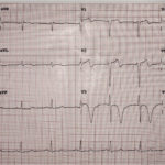
Initial electrocardiogram (ECG) revealed the classic biphasic T waves in V2 and V3 of Wellen’s syndrome (see red outlines). A second EKG demonstrated an evolving deeply inverted T wave (see blue outlines).

Discussion:

Wellen’s syndrome refers to specific ECG abnormalities in the precordial T wave segments of V2-V3.1,2 Recognition of this classic ECG pattern is important because it is highly specific for critical stenosis of the proximal left anterior descending artery.2,3 There are two patterns that can be seen in Wellen’s syndrome. Type A has deeply and symmetrically inverted T waves in V2-V3 (seen in 75% of cases) and Type B has biphasic T waves, with initially positive and terminally negative T waves in V2-V3 (seen in 25% of cases).4 The T waves can eventually evolve from type B to type A, which occurred in this patient while he was in the emergency department.

Clinically, patients may be pain-free when the pattern is detected. They may have normally or minimally elevated cardiac enzymes. Wellen’s syndrome can be the pre-infarction stage of coronary artery disease, meaning that these patients are at high risk for extensive anterior wall myocardial infarction within days to weeks.3 This patient was taken to the catheterization lab.  Angiogram revealed a subtotal proximal left anterior descending artery occlusion for which a drug-eluting stent was placed and the patient was discharged in good condition.

Topics:

Wellen’s, STEMI, electrocardiogram, ECG, coronary artery disease, cardiology.

References:

  1. Kyaw K, Latt H, Aung SS, Tun TM, Phoo WY, Yin HH. Atypical presentation of acute coronary syndrome and importance of Wellens’ syndrome. Am J Case Rep. 2018;19:199-202. doi: 10/12659/AJCR.907992
  2. Nisbet BC, Zlupko G. Repeat Wellen’s syndrome: case report of critical proximal left anterior descending artery restenosis. J Emerg Med. 2010;39(3)305-08. doi: 10.1016/j.jemergmed.2007.10.043
  3. Rosales-Castillo A, Plaza-Carrera J. Wellen’s syndrome. Rev Clin Esp. 2017;217(8):491.
  4. Hsu YC, Hsu CW, Chen TC. Type B Wellen’s syndrome: Electrocardiogram patterns that clinicians should be aware of. Ci Ji Yi Xue Za Zhi. 2017;29(2):127-128. doi: 10.4103/tcmj.tcmj_26_17
Icon

Wellens' Syndrome - Case Report

1 file(s) 476 KB
Download
Icon

Wellens' Syndrome - Images

1 file(s) 5.2 MB
Download
ECGIssue 4:1

Reviews:

No ratings yet.

Please rate this


Arteriovenous Graft Pseudoaneurysm

29 Dec, 18

Pericardial Clot on Point-of-Care Ultrasound

29 Dec, 18
JETem is an online, open access, peer-reviewed journal-repository for EM educators

Most Viewed

  • Telemedicine Consult for Shortness of Breath Due to Sympathetic Crashing Acute Pulmonary Edema
  • Anticholinergic Toxicity in the Emergency Department
  • The Suicidal Patient in the Emergency Department Team-Based Learning Activity
  • Child Maltreatment Education: Utilizing an Escape Room Activity to Engage Learners on a Sensitive Topic
  • Acute Chest Syndrome

Visit Our Collaborators

About

Education

Learners should benefit from active learning. JETem accepts submissions of team-based learning, small group learning, simulation, podcasts, lectures, innovations, curricula, question sets, and visualEM.

Scholarship

We believe educators should advance through the scholarship of their educational work. JETem gives educators the opportunity to publish scholarly academic work so that it may be widely distributed, thereby increasing the significance of their results.

Links

  • Home
  • Aim and Scope
  • Current Issue
  • For Reviewers
  • Instructions for Authors
  • Contact Us

Newsletter

Sign up to receive updates from JETem regarding newly published issues and findings.

Copyright © 2016 JETem. All rights reserved.