Issue 8:4
Low-Cost Fishhook Removal Simulation
DOI: https://doi.org/10.21980/J8Q64PThe goal of this small group session is to fill the gap in training on fishhook injuries. At the end of the session participants should be able to describe the parts of a fishhook, as well as demonstrate and have increased confidence in performing multiple fishhook removal techniques.
Enneagram in EM
DOI: https://doi.org/10.21980/J8ZM0GBy the end of this session, the learner will be able to: 1) Self-identify with a primary enneagram personality type. 2) List the fears, desires, and motivations of the enneagram type. 3) Describe struggles in interacting with other disparate enneagram types. 4) Discuss strategies for success in facing conflict and interacting with other team members.
Adolescent with Diabetic Ketoacidosis, Hypothermia and Pneumomediastinum
DOI: https://doi.org/10.21980/J8FP8JBy the end of the simulation, learners will be able to: 1) develop a differential diagnosis for an adolescent who presents obtunded with shortness of breath; 2) discuss the management of diabetic ketoacidosis; 3) discuss management of hypothermia in a pediatric patient; 4) discuss appropriate ventilator settings in a patient with diabetic ketoacidosis; and 5) demonstrate interpersonal communication with family, nursing, and consultants during high stress situations.
Ventricular Tachycardia
DOI: https://doi.org/10.21980/J8KD2RAt the conclusion of the simulation session, learners will be able to: 1) identify the different etiologies of VT, including structural heart disease, acute ischemia, and acquired or congenital QT syndrome; 2) describe confounding factors of VT, such as electrolyte abnormalities and sympathetic surge; 3) describe how to troubleshoot an unsuccessful synchronized cardioversion, including checking equipment connections, increasing delivered energy, and changing pad placement; 4) compare and contrast treatments of VT based on suspected underlying etiology; 5) describe reasons to activate the cardiac catheterization lab other than occlusive myocardial infarction; and 6) identify appropriate disposition of the patient to the cardiac catheterization lab.
Inhalational Injury Secondary to House Fire
DOI: https://doi.org/10.21980/J8TW7NAt the conclusion of the simulation session, learners will be able to: 1) recognize the indications for intubation in a thermal burn/inhalation injury patient; 2) develop a systematic approach to an inhalational injury airway; and 3) recognize indications for transfer to burn center.
Little Patients, Big Tasks – A Pediatric Emergency Medicine Escape Room
DOI: https://doi.org/10.21980/J89W70By the end of this small group exercise, learners will be able to: 1) demonstrate appropriate dosing of pediatric code and resuscitation medications; 2) recognize normal pediatric vital signs by age; 3) demonstrate appropriate use of formulas to calculate pediatric equipment sizes and insertion depths; 4) recognize classic pediatric murmurs; 5) appropriately diagnose congenital cardiac conditions; 6) recognize abnormal pediatric electrocardiograms (ECGs); 7) identify life-threatening pediatric conditions; 8) demonstrate intraosseous line (IO) insertion on a pediatric model; and 9) demonstrate appropriate use of the Neonatal Resuscitation Protocol (NRP®) algorithms.
Point-Of-Care Ultrasound Use for Detection of Multiple Metallic Foreign Body Ingestion in the Pediatric Emergency Department: A Case Report
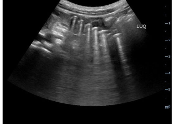
DOI: https://doi.org/10.21980/J83D2DBedside POCUS was performed on the patient’s abdomen using the curvilinear probe. The left upper quadrant POCUS image demonstrates multiple hyperechoic spherical objects with shadowing and reverberation artifacts concerning multiple foreign body ingestions. Though the patient and mother initially denied knowledge of foreign body ingestion, on repeated questioning after POCUS findings, the patient admitted to his mother that he ate the spherical magnets he received for his birthday about one week ago. The patient swallowed these over the course of two days. The presence of multiple radiopaque foreign bodies was confirmed with an abdominal X-ray.
Sonographic Retrobulbar Spot Sign in Diagnosis of Central Retinal Artery Occlusion: A Case Report
DOI: https://doi.org/10.21980/J8735PThe bedside ocular ultrasound (B-scan) was significant for small, hyperechoic signal (white arrow) in the distal aspect of the optic nerve, concerning for embolus in the central retinal artery. Subsequent direct fundoscopic exam was significant for a pale macula with cherry red spot (black arrow), consistent with central retinal artery occlusion (CRAO).


