A Case Report of Right Atrial Thrombosis Complicated by Multiple Pulmonary Emboli: POCUS For the Win!
ABSTRACT:
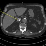
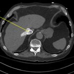
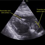
A 78-year-old gentleman presented to the emergency department (ED) for palpitations and dizziness. He had a complicated medical history including atrial fibrillation (AF), recently status post a Watchman procedure, oxygen-dependent chronic obstructive pulmonary disease (COPD), and heart failure with preserved ejection fraction (HFpEF). Point-of-care ultrasound (POCUS) revealed the presence of an intracardiac right atrial thrombus. Computed tomography (CT) angiography confirmed the presence of multiple pulmonary emboli (PE), and extension of the thrombus into the inferior vena cava. Pulmonary emboli are a common complication of thrombus in the right atrium. Management may include anticoagulation, thrombolysis, or thrombectomy. This case highlights that emergency physicians can expedite the diagnosis of intracardiac thrombus by using POCUS. The case presented describes a medically complex patient presenting with symptomatic right intracardiac and inferior vena caval thrombosis complicated by multiple PE. Point-of care ultrasound of the heart and lungs were included in his initial assessment, revealing findings of an intracardiac thrombus, and ruling out multiple other differential diagnoses including pericardial tamponade, pleural effusion, pulmonary edema, and pneumothorax. This finding changed the trajectory of this patient’s evaluation and management, and demonstrates the important role of POCUS in the care of ED patients with undifferentiated cardiopulmonary symptoms.
Topics:
Point-of care ultrasound (POCUS), focused cardiac ultrasound (FOCUS), inferior vena cava thrombosis, right atrial thrombosis, pulmonary embolism, computed tomography, echocardiography.


