Pericardial Clot on Point-of-Care Ultrasound
History of present illness:
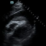
A 22-year-old female was brought to the emergency department (ED) by ambulance after sustaining multiple stab wounds to the chest, abdomen and flank. Upon initial evaluation, she was alert and oriented, in moderate distress due to pain but was verbally responsive. Vitals included a heart rate of 94 beats per minute with palpable radial pulses but no auscultable blood pressure. Patient was also noted to have pale and cool extremities. Bedside ultrasound exam performed immediately upon arrival showed a large pericardial effusion with tamponade physiology and a pericardial clot.
Significant findings:
Focused assessment with sonography in trauma (FAST) scan was positive for a clinically significant pericardial effusion as evidenced by the hypoechoic fluid around the myocardium, indicated by the blue arrow in image 2. Findings are also consistent with tamponade process as evidenced by restricted expansion and collapse of the right ventricle during diastole. The hyperechoic floating structure between the pericardium and myocardium, adjacent to the right ventricle, represents a pericardial clot, indicated by the white arrow.The density of the pericardial clot differs from that of the myocardium, thus serving as an additional variable to avoid confusing this as part of the myocardial structure.
Discussion:
As traumatic pericardial tamponade can quickly lead to hemodynamic collapse. Advanced trauma life support (ATLS) guidelines recommend the use of point-of-care ultrasound immediately following the primary survey to evaluate for free fluid in the pericardial sac. The FAST scan is both sensitive (96.8%) and specific (96.9%) for the detection of pericardial effusion in the context of trauma.1-2 In the presence of a negative FAST scan, a high index of suspicion should be maintained for pericardial effusion if large hemothoraces are present because sensitivity in these cases can be as low as 20%.3 The reason for this may be two-fold. First, a laceration in the pericardium leads to blood decompressing into the pleural cavity, resulting in an empty pericardial sac and an associated hemothorax. The second, fluid in the pleural sac obscures ultrasound interpretation by altering the echogenicity of the pericardial sac and the pleural space. This leads to potentially masking an underlying pericardial effusion.3 The importance of the FAST scan is also highlighted by the fact that electrocardiogram findings are often nonspecific and that only 10%-40% of tamponade cause the classic Beck’s triad of hypotension, jugular venous distension, and muffled heart sounds.4
The gold standard for the management of cardiac tamponade is decompression via emergent pericardiocentesis. In the traumatic setting, however, clotted blood may be present. Aspiration of clotted hemopericardium is often not possible and surgical drainage through either subxiphoid pericardiotomy or thoracotomy is warranted.5-6 Pericardiotomy in recent studies has been found to be superior to pericardiocentesis for a multitude of reasons, including decreased rates of failure to relieve pressure, decreased rates of complications such as damage to surrounding structures, and increased rates of survival.7
In our patient, the use of point-of-care ultrasound was able to quickly make the diagnosis of cardiac tamponade and clotted blood in the pericardium. Knowing this information allowed this patient to undergo definitive management. In this case, the patient had large bore peripheral intravenous access established and was taken emergently to the operating room for a thoracotomy. This revealed a left ventricle stab wound, inferior vena cava through-and-through stab wound, pericardial effusion with tamponade and clotted blood. A pericardiectomy was performed and the patient eventually stabilized.
Topics:
Pericardial Clot, Cardiac Tamponade, point-of-care ultrasound, pericardiotomy, hemopericardium.
References:
- Richards JR, McGahan JP. Focused assessment with sonography in trauma (FAST) in 2017: what radiologists can learn. Radiology. 2017; 283: 30-48. doi: 10.1148/radiol.2017160107
- Rozycki GS, Feliciano DV, Ochsner GM, et al. The role of ultrasound in patients with possible penetrating cardiac wounds: a prospective multicenter study. J Trauma. 1999; 46:543–552.
- Meyer DM, Jessen ME, Grayburn PA. Use of echocardiography to detect occult cardiac injury after penetrating thoracic trauma: a prospective study. J Trauma. 1995; 39:902-909.
- Guberman BA, Fowler NO, Engel PJ, Gueron M, Allen JM. Cardiac tamponade in medical patients. Circulation. 1981;64: 633–640.
- Ishida K, Kinoshita Y, Iwasa N, et al. Emergency room thoracotomy for acute traumatic cardiac tamponade caused by a blunt cardiac injury: a case report. Int J Surg Case Rep. 2017; 35: 21-24. doi: 10.1016/j.ijscr.2017.03.009
- Kurimoto Y, Hase M, Nara S, et al. Blind subxiphoid pericardiotomy for cardiac tamponade because of acute hemopericardium. J Trauma. 2006; 61(3): 582-585.
- Kurimoto Y, Maekawa K, Tanno K, et al. Blind subxiphoid pericardiotomy to relieve critical acute hemopericardium: a final report. Eur J Trauma Emerg Surg. 2012; 38(5): 563-568. doi: 10.1007/s00068-012-0200-3


