Pulmonary Embolism: Diagnosis by Computerized Tomography without Intravenous Contrast
History of present illness:
A 91-year-old female with a history of deep venous thrombosis presented to a rural Emergency Department with symptoms of dyspnea and chest pain radiating towards her back. She was not on anticoagulation secondary to fall concerns. A chest radiograph revealed only a widened mediastinum. Intravenous contrast could not be administered secondary to decreased renal function; ventilation perfusion scanning was not available. A CT scan of the chest was performed without contrast to evaluate the patient’s dyspnea and widened mediastinum.
Significant findings:
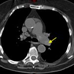
Non-contrast CT of the chest demonstrates hyper-densities within both central and sub-segmental pulmonary arteries bilaterally (see yellow arrows). The right ventricle is dilated.
Discussion:
The diagnosis of pulmonary embolism is usually made by visualizing intravenous contrast filling defects within the pulmonary arteries on CT angiography of the chest. Ventilation perfusion scanning is an alternative modality, but was not available in this case. A hyper-dense lumen sign on non-contrast chest CT1 can identify pulmonary emboli with a reported sensitivity of 36%.2
Utilizing non-contrasted CT of the chest to identify hemodynamically significant central thrombi when IV contrast is not an option may allow for initiation of therapy in a timely manner or may help identify PE when it may not be the primary consideration.
Topics:
Pulmonary embolism, respiratory, PE, CT, pulmonology.
References:
- Kanne JP, Gotway MB, Thoongsuwan N, Stern EJ. Six cases of acute central pulmonary embolism revealed on unenhanced multidetector CT of the chest. AJR Am J Roentgenol. 2003;180:1661-1664. doi: 10.2214/ajr.180.6.1801661
- Tatco VR, Piedad HH. The validity of hyperdense lumen sign in non-contrast chest CT scans in the detection of pulmonary thromboembolism. Int J Cardiovasc Imaging. 2011;27:433-440. doi: 10.1007/s10554-010-9673-5


