Latest Articles
Open Chest Wound with Sternal Fracture in the Emergency Department, a Case Report
DOI: https://doi.org/10.5070/M5.52202The image demonstrates the large chronic-appearing wound of the patient’s anterior chest as well as the visible fractured segments of the patient’s exposed sternum. The sternum is necrotic appearing concerning for a chronic process including osteomyelitis and malignancy. Purulent drainage is visible on the wound consistent with an infectious process.
Effects of Volume Overload: A Case Report of an Edema Bulla
DOI: https://doi.org/10.5070/M5.52206This image shows a large edema bulla on the patient's right shin. The bulla is 10 x 10 cm, filled with serous fluid, has a spontaneously occurring defect in the skin of the superior portion of the bulla, and is non-erythematous. The bulla is much larger than the 1-5 cm edema bullae described in the literature. As edema bulla is primarily a clinical diagnosis, taking the full history and physical exam into account is essential to recognize these bullae.
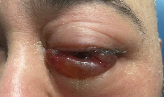
A Case Report of Carotid Cavernous Fistula: A Commonly Missed Diagnosis
DOI: https://doi.org/10.5070/M5.52242The initial physical exam performed by the ED provider revealed severe left eye chemosis, clear drainage, visual acuity of right eye 20/100 and left eye 20/400, and a left eye IOP of 52. There was a deficit of extraocular movement in all directions of gaze and limitation in all visual fields in the left eye. The MRI showed that at the level of the eye, the left cavernous sinus is asymmetrically enlarged compared to the right (red arrow) with an enlarged left inferior petrosal sinus with internal flow void on the pre-contrast MRI images (blue arrow). The orange arrow notes a central filling defect of the left superior ophthalmic vein on the MRA.


