• Registration
  • Login
JETem
  • Home
  • About
    • Aim and Scope
    • Our Team
    • Editorial Board
    • FAQ
  • Issues
    • Current Issue
    • Ahead of Print
    • Past Issues
  • Visual EM
    • Latest Visual EM
    • Search Visual EM
    • Thumbnail Library
  • For Authors
    • Instructions for Authors
    • Submit to JETem
    • Photo Consent
    • Policies
      • Peer Review Policy
      • Copyright Policy
      • Editorial Policy, Ethics and Responsibilities
      • Conflicts of Interest & Informed Consent
      • Open Access Policy
  • For Reviewers
    • Instructions for JETem Reviewers
    • Interested in Being a JETem Reviewer?
  • Topic
    • Abdominal / Gastroenterology
    • Administration
    • Board Review
    • Cardiology / Vascular
    • Clinical Informatics, Telehealth and Technology
    • Dermatology
    • EMS
    • Endocrine
    • ENT
    • Faculty Development
    • Genitourinary
    • Geriatrics
    • Hematology / Oncology
    • Infectious Disease
    • Miscellaneous
    • Neurology
    • Ob / Gyn
    • Ophthalmology
    • Orthopedics
    • Pediatrics
    • Pharmacology
    • Procedures
    • Psychiatry
    • Renal / Electrolytes
    • Respiratory
    • Social Determinants of Health
    • Toxicology
    • Trauma
    • Ultrasound
    • Urology
    • Wellness
    • Wilderness
  • Modality
    • Curricula
    • Innovations
    • Lectures
    • Oral Boards
      • Structured Interview
      • Communication Case
    • Podcasts
    • Simulation
    • Small Group Learning
    • Team Based Learning
    • Visual EM
  • Contact Us

A Case of Otomastoiditis

Chad Correa, BS* and Claire Thomas, MD^

*University of California, Riverside, School of Medicine, Riverside, CA
^University of California, Irvine, Department ofEmergency Medicine, Orange, CA

Correspondence should be addressed to Chad Correa, BS at chad.correa@medsch.ucr.edu 

DOI: https://doi.org/10.21980/J8RK89Issue 3:2
ENTInfectious DiseaseVisual EM
No ratings yet.
Creative Commons images

History of present illness:

A 45-year-old male presented to the emergency department with chief complaint of left ear pain with yellow discharge for several days. He had associated decrease in appetite. The patient denied any history of fever, headache, dizziness, hearing loss, or tinnitus.  On physical exam, the patient was found to have yellow discharge in the left ear canal with a perforated tympanic membrane.  He was also found to have significant tenderness to palpation over the left mastoid bone.

Significant findings:

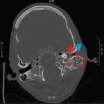
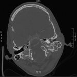
The patient underwent computed tomography (CT) of the head which revealed opacification of the left middle ear (red arrow) and mastoid air cells (red circles). Additionally, there was thickening of the soft tissues of the external auditory canal (blue arrowhead), likely reflecting concurrent otitis externa.  Based on the imaging, he was admitted for findings consistent with acute otomastoiditis.

Discussion:

Mastoiditis is an infection of the mastoid air cells of the temporal bone often secondary to undertreated or untreated otitis media.  On CT, purulent material and mucosal swelling cause opacification of the air cells, as seen in this patient.1 The inflammation can erode into the mastoid septae causing acute coalescent otomastoiditis.  Mastoiditis can invade intracranially leading to sigmoid sinus thrombosis, perisinus empyema, epidural abscess, subdural abscess or meningitis.1

Presentation of mastoiditis includes fever, otalgia and otorrhea.  Patients may also have erythema and swelling overlying the mastoid process.2   Most patients presenting with acute mastoiditis will require hospital admission for intravenous (IV) antibiotics.  Antibiotics should cover the common causative agents of otitis media including H influenzae, M catarrhalis, S pneumoniae as well as S aureus. Mastoidectomy is reserved for cases where medical management fails.3

In the antibiotic era, the incidence of clinically significant acute mastoiditis has decreased to 1.2-2 per 100,000.2 However, it is important to recognize that acute mastoiditis may present with severe intracranial complications resulting in significant morbidity and mortality.3   Therefore, patients with suspected mastoiditis should receive a thorough physical examand cranial imaging studies to evaluate for bone erosion and other sequelae.4

The patient in this case remained afebrile without leukocytosis throughout hospitalization.  A final blood culture yielded gram positive cocci, gram negative cocci, gram positive rods and methicillin resistant staph aureus with Clindamycin susceptibility. The otolaryngologist (ENT) started the patient on IV ceftriaxone and IV clindamycin for 72 hours. He was discharged with ENT follow-up on an oral regimen of clindamycin and cefuroxime for two weeks. He was also administered ofloxacin otic solution throughout his hospitalization and at discharge.

Topics:

Mastoiditis, otomastoiditis, otitis externa, CT

References:

  1. Patel KM, Almutairi A, Mafee MF. Acute otomastoiditis and its complications: role of imaging. Operative Techniques in Otolaryngology. 2014;25(1):21-28. doi: 10.1016/j.otot.2013.11.004
  2. Rubin MA, Ford LC, Gonzales R. Sore throat, earache, and upper respiratory symptoms. In: Kasper DL, Hauser SL, Jameson JL, Fauci AS, Longo DL, and Loscalzo J, eds. Harrison’s Principles of Internal Medicine. 19th ed. New York, NY: McGraw-Hill; 2014:247-254.
  3. Jauch EC, White DR, Knoop KJ. Ear, nose, and throat conditions. In: Knoop KJ, Stack LB, Storrow AB, and Thurman RJ, eds. The Atlas of Emergency Medicine. 4th ed. New York, NY: McGraw-Hill; 2016:99-134.
  4. Luntz M, Bartal K, Brodsky A, Shihada R. Acute mastoiditis: the role of imaging for identifying intracranial complications. The Laryngoscope. 2012;122(12):2813-2817. doi: 10.1002/lary.22193
Icon

Left Mastoiditis - Case Report

1 file(s) 531 KB
Download
Icon

Left Mastoiditis - Images

1 file(s) 1.5 MB
Download
CTIssue 3:2

Reviews:

No ratings yet.

Please rate this


Creative Commons images

Osborn Waves in a Severely Hypothermic Patient

13 Apr, 18
Creative Commons images

Glass Foreign Body Hand Radiograph

13 Apr, 18

JETem is an online, open access, peer-reviewed, journal-repository for EM educators. We are PMC Indexed.

Most Viewed

  • The Silent Saboteur: Teaching the Clinical Implications of Occult Hypoxemia & Social Determinants of Health via a Pulmonary Embolism Case
  • Diabetic Ketoacidosis and Necrotizing Soft Tissue Infection
  • My Broken Heart
  • Stabilization of Cardiogenic Shock for Critical Care Transport, a Simulation
  • Innovative Ultrasound-Guided Erector Spinae Plane Nerve Block Model for Training Emergency Medicine Physicians

Visit Our Collaborators

Creative Commons Licence
This work is licensed under a Creative Commons Attribution 4.0 International License.

About

Education

Learners should benefit from active learning. JETem accepts submissions of team-based learning, small group learning, simulation, podcasts, lectures, innovations, curricula, question sets, and visualEM.

Scholarship

We believe educators should advance through the scholarship of their educational work. JETem gives educators the opportunity to publish scholarly academic work so that it may be widely distributed, thereby increasing the significance of their results.

Links

  • Home
  • Aim and Scope
  • Current Issue
  • For Reviewers
  • Instructions for Authors
  • Contact Us

Newsletter

Sign up to receive updates from JETem regarding newly published issues and findings.

Copyright Creative Commons Attribution 4.0 International